Encyclopedia entry
Auriculotemporal Nerve
Abstract
This nerve carries the otic ganglion, which is derived from glossopharyngeal neurons. The nerve usually arises by two roots from the posterior division of the mandibular nerve. The two roots usually surround the middle meningeal nerve before joining to form a single trunk. A variation in this relationship has been described in which the middle meningeal artery pierces the anterior root instead of passing between the two roots. According to Baumel, Vanderheiden and McElenney the auriculotemporal nerve is commonly misrepresented in both illustrations and textbooks.
This nerve carries the otic ganglion, which is derived from glossopharyngeal neurons.
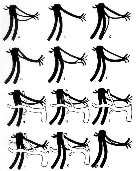
The nerve usually arises by two roots from the posterior division of the mandibular nerve. The two roots usually surround the middle meningeal nerve before joining to form a single trunk. A variation in this relationship has been described in which the middle meningeal artery pierces the anterior root instead of passing between the two roots.
According to Baumel, Vanderheiden and McElenney the auriculotemporal nerve is commonly misrepresented in both illustrations and textbooks. Their 85 dissections of the nerve demonstrate that the roots of the "typical" auriculotemporal nerve do not form a tight buttonhole around the middle meningeal artery.
Instead the roots outline an elongated, V-shaped interval with the roots widely separated from one another. At their junction the roots form a short trunk which immediately breaks up in line with the posterior border of the mandible into a spray of branches. The superficial temporal ramus of the auriculotemporal nerve should not be considered as the main continuation of the nerve, but merely as its largest branch. A substantial portion of the nerve makes up its two communicating rami with the facial nerve; these are the strongest and most consistent of the many peripheral communications between trigeminal and facial nerves. Common variations in configuration, branching, and relationships of the nerve are discussed in their paper.
Anatomy Atlases. Image hosted by the site it comes from. Open the original page.
Riferimenti dell’opera originale
Elenco riportato come compare nella fonte, senza normalizzazione dei formati bibliografici.
- Baumel, J.J. and D.Y. Beard (1961) The accessory meningeal artery of man. J. Anat. 95:386-402.
- Baumel, J.J., Vanderheiden, J.P. and J. E. McElenney. (1971) The auriculotemporal Nerve in Man. Am. J. Anat. 130(4):431-440.
- Cushing, H. (1904) The sensory distribution of the fifth cranial nerve. Johns Hopkins Hospital Bulletin 15:213-232.
- Dixon, A.F. (1896) On the development of the branches of the fifth cranial nerve in man. Trans. Roy. Scient. Soc., Dublin. 6(S.2): 19-69.
- Huber, G.C. (1930) Piersol's Human Anatomy, 9th ed., L.B. Lippincott Co., Philadelphia.
- Krizan, Z. (1956) Beitrag zur Kenntnis des dritten Trigeminusastes. Pract. oto-rhino-laryng. 18:144-154.
- Plá Majó, B. (1934) Contribucion al estudio del ojal nervioso del auriculo-temporal. Arquivo Anat. Antrop. 16:233-244.
- Schaefer, E.A., Symington, J. and T.H. Bryce., Eds. (1915) Quain's Anatomy, 11th ed. Longmans, Green, and Co., London.
- Weigner, C. (1898) Le ganglion otique. Bibliog. Anat. 6:302-306.
Anatomical classification
- Systems:
- Nervous system
Content provenance
Adaptation
Adaptation of «Auriculotemporal Nerve — Illustrated Encyclopedia of Human Anatomic Variation: Opus I: Muscular System» by Ronald A. Bergman, Adel K. Afifi, Ryosuke Miyauchi. Source: https://www.anatomyatlases.org/AnatomicVariants/NervousSystem/Text/AuriculotemporalNerve.shtml Original work distributed under the CC BY-NC-SA 4.0 licence (https://creativecommons.org/licenses/by-nc-sa/4.0/deed.it). Adaptation by Redazione AIVA. Changes from the original: Testo originale inglese riprodotto integralmente e riorganizzato nel formato scheda del portale (sintesi, testo, riferimenti), senza modificarne le affermazioni. La traduzione italiana non è ancora stata eseguita, e la pagina lo dichiara. Le figure non sono state copiate: sono collegate alla pagina d’origine. This publication is not approved or endorsed by the authors of the original work.
Licence
This content is distributed under the licence CC BY-NC-SA 4.0. Licences and attribution