Encyclopedia entry
Common Iliac Artery
Abstract
The common iliac arteries are as short as 1.2 cm or as long as 11 cm; the usual length is 3.7-7.5 cm in 72% of individuals studied. The remaining 28% were divided equally, being longer or shorter than usual length.These variations in length may depend on whether the aorta bifurcates above or below the usual point, or on whether the common iliac arteries divide higher or lower than usual. A low aortic bifurcation is more common than a high bifurcation, as is also the case with the common iliacs. The bifurcation of the common iliacs varies.
The common iliac arteries are as short as 1.2 cm or as long as 11 cm; the usual length is 3.7-7.5 cm in 72% of individuals studied. The remaining 28% were divided equally, being longer or shorter than usual length.These variations in length may depend on whether the aorta bifurcates above or below the usual point, or on whether the common iliac arteries divide higher or lower than usual. A low aortic bifurcation is more common than a high bifurcation, as is also the case with the common iliacs.
The bifurcation of the common iliacs varies. In 68% of individuals studied, the division occurs between the fifth lumbar vertebra and the upper border of the sacrum. Approximately 12% occur above the fifth vertebra and 17% below the upper border of the sacrum. More frequently, the left common iliac artery divides further distally than the right.
The common iliacs may be absent; the external and internal iliacs then arise together from the distal end of the aorta.
Either artery may provide a large branch, such as the iliolumbar or the lateral sacral or middle sacral, sometimes a lumbar, or occasionally an "accessory" renal or a gonadal artery. In some cases, the right and left common iliacs are joined by a transverse anastomotic vessel.
The embryonic axis (ischiadic or sciatic) artery may persist and be of significant size and importance to give rise to the popliteal artery.
Occasionally the common iliac fails to to bifurcate, sinking into the true pelvis to provide the usual pelvic branches and then emerging to continue as the external iliac.
Unusual branches of the common iliac include a middle colic, umbilical, obturator, and the circumflex iliac.
The absence of the common iliac artery was noted on the left side of a dissected body by Walsham (he also noted that horses do not have common iliac arteries). The aorta , in the case of Walsham's subject, divided into three branches opposite the middle of the fourth lumbar vertebra - into a right common iliac, a left external, and a left internal iliac. Cruveilhier reported one instance of the absence of the right common iliac artery.
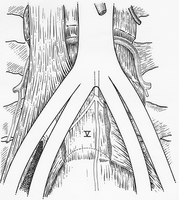
Middle Colic
Anatomy Atlases. Image hosted by the site it comes from. Open the original page.
Anatomy Atlases. Image hosted by the site it comes from. Open the original page.
Riferimenti dell’opera originale
Elenco riportato come compare nella fonte, senza normalizzazione dei formati bibliografici.
- Anson, B.J., Ed. (1966) Morris'Human Anatomy, 12th ed., The Blakiston Division, McGraw-Hill Book Company.
- Bergman, R.A., Thompson, S.A., Afifi, A.K. and F.A. Saadeh. (1988) Compendium of Human Anatomic Variation:Catalog, Atlas and World Literature. Urban & Schwarzenberg, Baltimore and Munich.
- Blair, Jr., C.B. and K. Nandy. (1965) Persistence of the axis artery of the lower limb. Anat. Rec. 152:161-172.
- Dufour, -. (1852) Trois artères vent se jeter dans le rein; une venant de l'aorte, et les deux autres de l'iliaque primitive. Bulletins et Mem. de la Société Anatomique de Paris XXVII(1):27.
- Gariraldi, G. (1876) Casi di anomalia nel decorso delle vene iliache primitive e della vena cava ascendente. Spallanzani 14:362-367.
- Gruber, W. (1880) Duplicität der Arteria spermatica interna bei Ursprung der supernumerären Arteria aus der Arteria iliaca communis. Arch. Pathol. Anat. Physiol. Klin. Med. 81:457-459.
- Hamann, C.A. (1892) Common iliac artery. University Medical Magazine, Philadelphia 5:185.
- Henle, J. (1868) Handbuch der Systematischen Anatomie des Menschen, Von Friedrich Vieweg und Sohn. Braunschweig.
- Latarjet, A. (1948) Testut's Traite d' Anatomie Humaine, 9th ed., G. Doin & Cie., Paris.
- Levi, G. (1902) Observations sur les variations des artères iliaques. Arch. Ital. Biol. 37:489.
- Levi, G. (1902) Morfologia delle arterie ilache. Arch. Ital. Anat. Embriol. 1:120-172, 295-346, 523-605.
- Paterson, A.M. (1910) Obliteration of the left common, external, and internal iliac arteries. J. Anat. Physiol. 44:56.
- Poynter, C.W.M. (1922) Congenital anomalies of the arteries and veins of the human body with bibliography. University Studies of the University of Nebraska 22:1-106.
- Schaefer, E.A., Symington, J, and T.H. Bryce, Eds. (1915) Quain's Anatomy, 11th ed., Longmans, Green and Co., London.
- Shepherd, F.J. (1882) On some anatomical variations: Common iliac arteries. Annals of Anatomy and Surgery. 6;171.
- von Willer, P. (1917-18) Eine seltene Varietät der Arteria iliaca communis sinistra. Anat. Anz. 50:155-158.
- Winslow, R. (1883) A study of the malformations, variations, and anomalies of the circulatory apparatus in man. Annals of Anatomy and Surgery 7:94.
Anatomical classification
- Systems:
- Cardiovascular system
Content provenance
Adaptation
Adaptation of «Common Iliac Artery — Illustrated Encyclopedia of Human Anatomic Variation: Opus I: Muscular System» by Ronald A. Bergman, Adel K. Afifi, Ryosuke Miyauchi. Source: https://www.anatomyatlases.org/AnatomicVariants/Cardiovascular/Text/Arteries/CommonIliac.shtml Original work distributed under the CC BY-NC-SA 4.0 licence (https://creativecommons.org/licenses/by-nc-sa/4.0/deed.it). Adaptation by Redazione AIVA. Changes from the original: Testo originale inglese riprodotto integralmente e riorganizzato nel formato scheda del portale (sintesi, testo, riferimenti), senza modificarne le affermazioni. La traduzione italiana non è ancora stata eseguita, e la pagina lo dichiara. Le figure non sono state copiate: sono collegate alla pagina d’origine. This publication is not approved or endorsed by the authors of the original work.
Licence
This content is distributed under the licence CC BY-NC-SA 4.0. Licences and attribution