Encyclopedia entry
Ilioinguinal Nerve
Abstract
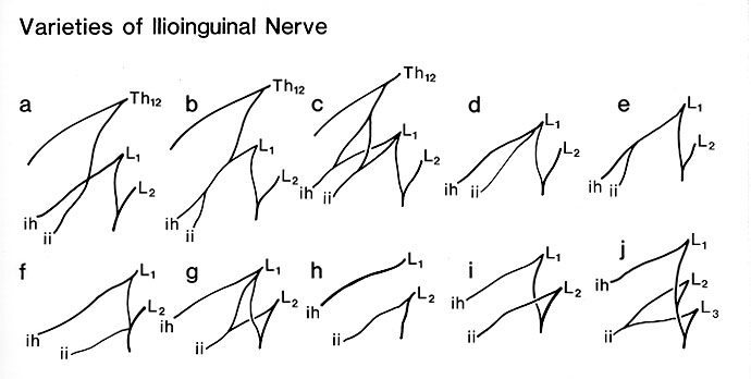
This nerve may be small and terminate near the iliac crest by joining the iliohypogastric nerve, which sends branches to replace the absent terminal part of the ilioinguinal nerve. When present, the nerve is replaced by either the genital (more commonly) or the femoral branches of the genitofemoral nerve. The ilioinguinal may provide a lateral cutaneous or iliac branch to supply the skin in the region of the anterior superior iliac spine. It may partially or completely replace the genital branch of the genitofemoral nerve or the lateral femoral cutaneous nerve.
This nerve may be small and terminate near the iliac crest by joining the iliohypogastric nerve, which sends branches to replace the absent terminal part of the ilioinguinal nerve.
When present, the nerve is replaced by either the genital (more commonly) or the femoral branches of the genitofemoral nerve.
The ilioinguinal may provide a lateral cutaneous or iliac branch to supply the skin in the region of the anterior superior iliac spine.
It may partially or completely replace the genital branch of the genitofemoral nerve or the lateral femoral cutaneous nerve.
The ilioinguinal and iliohypogastric nerves sometime arise as a common trunk, and in their course, usually separate between the transversus and internal oblique muscles.
The ilioinguinal nerve may be derived from the last thoracic nerve (T12), from a loop between the first and second lumbar nerves, or even from the second and third lumbar nerves. The rectus abdominis muscle may receive a branch from the ilioinguinal.
In one study of 200 bodies, the ilioinguinal nerve arose from the lumbar plexus in 72.5% and by a common trunk with the iliohypogastric nerve in 25%; it was absent in 2.5%. The ilioinguinal nerve was formed from one root in 92.5 % and from two roots in about 5% of cases. In 86%, the ilioinguinal carried fibers from one spinal nerve (primarily from L1), and in 11%, from two spinal nerves (T12, L1; L1, L2; or L2, L3).
Within the inguinal canal, the nerve usually lies ventral to the spermatic cord (60% of cases) but it may lie beneath (dorsal) the cord and/or within it.
This nerve may also leave the superficial inguinal ring at its lateral aspect instead of its usual medial position. In some cases, the nerve runs outside the inguinal ring.
Anatomy Atlases. Image hosted by the site it comes from. Open the original page.
Riferimenti dell’opera originale
Elenco riportato come compare nella fonte, senza normalizzazione dei formati bibliografici.
- Eckmann, I. (1976) Ein Beitrag zur Kenntnis des terminalen Verlaufs des N. ilioinguinalis. Anat. Anz. 140:15-30.
- Jamieson, R.W., Swigart, LaV.L. and B.J. Anson. (1952) Points of parietal perforation of the ilioinguinal and iliohypogastric nerves in relation to optimal sites for local anesthesia. Q. Bull. Northwestern University Medical School 26:22-26.
- Oelrich, T.M. and D.A. Moosman. (1977) The aberrant course of the cutaneous component of the ilioinguinal nerve. Anat. Rec. 189:233-236.
- Papadopoulos, N.J. and E.D. Katritsis. (1981) Some observations on the course and relations of the iliohypogastrics and ilioinguinal nerves (based on 348 specimens). Anat. Anz. 149:357-364.
- Zaluska, S. (1975) External structure of the ilioinguinal nerve in postnatal life in man. Folia Morphol. (Warsaw), 34:419-424.
Anatomical classification
- Systems:
- Nervous system
Content provenance
Adaptation
Adaptation of «Ilioinguinal Nerve — Illustrated Encyclopedia of Human Anatomic Variation: Opus I: Muscular System» by Ronald A. Bergman, Adel K. Afifi, Ryosuke Miyauchi. Source: https://www.anatomyatlases.org/AnatomicVariants/NervousSystem/Text/IlioinguinalNerve.shtml Original work distributed under the CC BY-NC-SA 4.0 licence (https://creativecommons.org/licenses/by-nc-sa/4.0/deed.it). Adaptation by Redazione AIVA. Changes from the original: Testo originale inglese riprodotto integralmente e riorganizzato nel formato scheda del portale (sintesi, testo, riferimenti), senza modificarne le affermazioni. La traduzione italiana non è ancora stata eseguita, e la pagina lo dichiara. Le figure non sono state copiate: sono collegate alla pagina d’origine. This publication is not approved or endorsed by the authors of the original work.
Licence
This content is distributed under the licence CC BY-NC-SA 4.0. Licences and attribution