Voce enciclopedica
Caviglia e piede: tarsali, metatarsali e falangi
Abstract
Le ossa del tarso sono variabili per dimensioni, forma e numero, con circa 30 ossicini tarsali accessori descritti. Anche metatarsali, falangi e numero delle dita possono essere aumentati o ridotti, e sono state riportate fusioni, duplicazioni e sesamoidi soprannumerari.
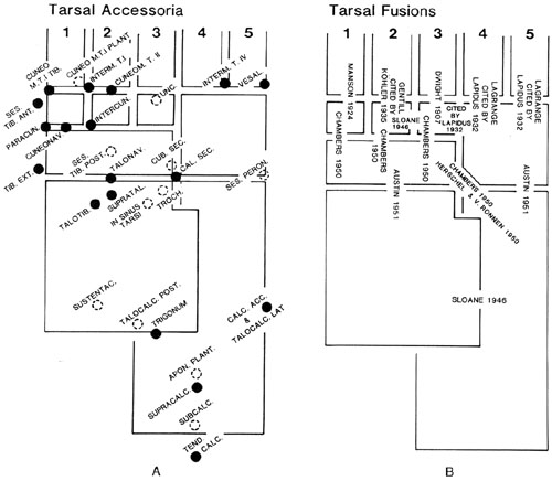
Le ossa del tarso sono variabili per dimensioni, forma e numero. Sono stati descritti circa 30 ossicini tarsali accessori. La diversità è stata messa in relazione in parte con la loro funzione nel carico ponderale. Le ossa del tarso sono comunemente ridotte a sei per la fusione del calcagno e dello scafoide.
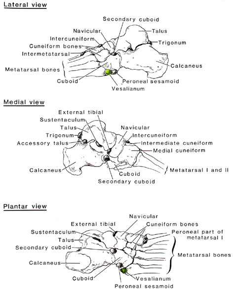
Nel tarso possono comparire elementi aggiuntivi rispetto ai sette normali. Il più frequente è la presenza di un osso triangolare (os trigonum), che può derivare dalla mancata fusione fra il tubercolo laterale del processo posteriore dell’astragalo e il centro di ossificazione primario dell’osso. La sua frequenza è riportata variamente al 2,5-11,4%. La variazione successiva per frequenza riguarda l’ossificazione indipendente della metà plantare e della metà dorsale del primo cuneiforme (os cuneiforme mediale bipartum). Possono trovarsi elementi aggiuntivi, probabilmente correlati a problemi di sviluppo del calcagno e dello scafoide. Alcuni di questi elementi aggiuntivi si trovano (a) in rapporto con il primo cuneiforme e il primo e secondo metatarsale (l’os intermetatarsum si riscontra nel 10% dei piedi), (b) sul lato mediale dell’articolazione cuneonavicolare (os paracuneiforme) e (c) fra il quinto metatarsale e il cuboide (os vesalianum). La tuberosità dello scafoide può costituire un osso separato (os tibiale externum). È stato descritto un cuboide secondario che origina dallo scafoide come una sporgenza che si separa dal lato laterale della superficie plantare dell’osso. All’estremità posteriore del sustentaculum può esservi un osso inconsueto detto os sustentaculi proprium. L’os calcaneum secundarium è un raro piccolo osso presente sul dorso fra calcagno, cuboide, scafoide e testa dell’astragalo.
Sul dorso, alle estremità prossimali del primo e del secondo cuneiforme, si può occasionalmente trovare un osso intercuneiforme libero (os intercuneiforme) nella fossa intercuneiforme.
La porzione laterale dello scafoide può essere unita al cuboide, risultando così separata dal resto dello scafoide. Ne consegue l’articolazione del cuboide con l’astragalo.
È stata riportata una sinostosi astragalo-scafoidea congenita. Una faccetta articolare aggiuntiva è stata osservata sulla superficie laterale del calcagno, anteriormente alla troclea peroneale, in circa il 7% dei casi.
Lo scafoide accessorio è il più grande osso soprannumerario del piede e l’incidenza riportata nei piedi normali è di circa il 10-21% (Kiter). Sono stati descritti tre tipi di scafoide accessorio (AN): il tipo 1, o tibiale externum, è un osso sesamoide di 2x3 mm di diametro incluso nella porzione distale del tendine del muscolo tibiale posteriore. L’AN di tipo 2 è un osso triangolare o a forma di cuore che può misurare fino a 12 mm. L’AN di tipo 3 è una tuberosità scafoidea prominente e si ritiene rappresenti un AN di tipo 2 fuso. I tipi 2 e 3 hanno importanza clinica (Kiter). Il tibiale posteriore può inserirsi direttamente su uno scafoide accessorio senza estendersi alla pianta del piede.
I metatarsali possono essere ridotti di numero a quattro elementi.
Ossa sesamoidi compaiono in articolazioni diverse dalla prima metatarsofalangea, ma sono più rare di quelle riscontrate nella mano. In uno studio su 246 piedi, Pfitzner trovò due sesamoidi nel quinto dito in circa il 6% e un sesamoide nel secondo dito in circa il 2%. Sesamoidi furono inoltre riscontrati nell’articolazione interfalangea dell’alluce nel 55% e nella stessa articolazione del secondo dito in circa l’1%.
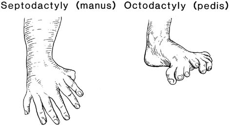
Le falangi possono essere aumentate o diminuite di numero e di dimensioni. Il numero delle dita può essere aumentato o diminuito. È stata riportata l’ottodattilia. Le dita possono essere fuse fra loro o assenti.
Una rassegna critica delle variazioni del tarso è stata fornita da R. O’Rahilly.
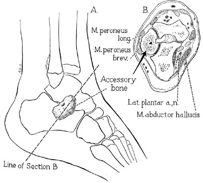
Un osso tarsale anomalo.
Anatomy Atlases. Immagine ospitata dal sito d’origine. Apri la pagina originale.
Settodattilia (manus) e ottodattilia (pedis).
Anatomy Atlases. Immagine ospitata dal sito d’origine. Apri la pagina originale.
Ossa tarsali accessorie. Fusioni tarsali.
Anatomy Atlases. Immagine ospitata dal sito d’origine. Apri la pagina originale.
Ossa tarsali soprannumerarie.
Anatomy Atlases. Immagine ospitata dal sito d’origine. Apri la pagina originale.
Duplicazione dell’alluce. Duplicazione del quinto dito del piede. Esadattilia (manus).
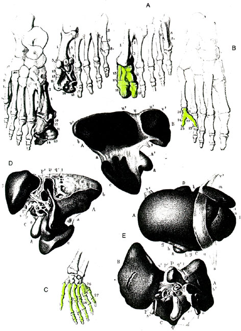
Anatomy Atlases. Immagine ospitata dal sito d’origine. Apri la pagina originale.
Polidattilia.
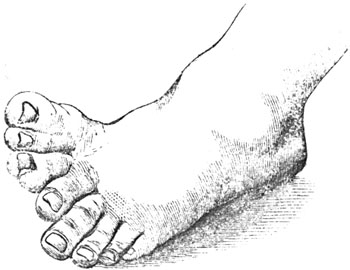
Anatomy Atlases. Immagine ospitata dal sito d’origine. Apri la pagina originale.
Os vesalianum.
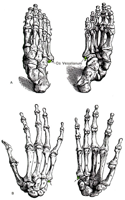
Anatomy Atlases. Immagine ospitata dal sito d’origine. Apri la pagina originale.
Duplicazione dell’alluce.
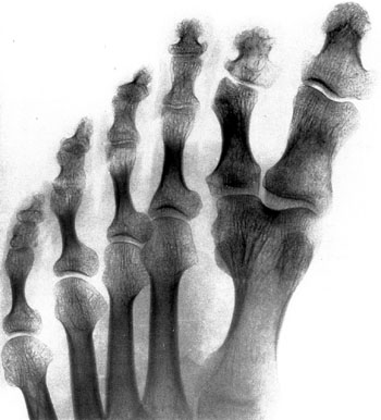
Anatomy Atlases. Immagine ospitata dal sito d’origine. Apri la pagina originale.
Ossa tarsali soprannumerarie.
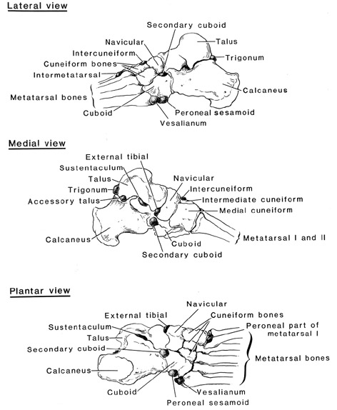
Anatomy Atlases. Immagine ospitata dal sito d’origine. Apri la pagina originale.
Duplicazione del quinto dito del piede da quinto metatarsale bifido.
Sdoppiamento dell’alluce, parte 1 di 2
Sdoppiamento del dito, parte 2 di 2
Trigonum, parte 1 di 8
Trigonum, parte 2 di 8
Trigonum, parte 3 di 8
Trigonum, parte 4 di 8
Trigonum, parte 5 di 8
Trigonum, parte 6 di 8
Trigonum, parte 7 di 8
Trigonum, parte 8 di 8
Riferimenti dell’opera originale
Elenco riportato come compare nella fonte, senza normalizzazione dei formati bibliografici. Le voci non sono tradotte: un riferimento bibliografico si cita nella lingua in cui è pubblicato.
- Aitken, D.M. (1905) A note on the variations of the tibia and astragalus. J. Anat. Physiol. 39:489-491.
- Anonymous. (1922) Anatomie du Vivant. Septodactylie (manus) et Octodactylie (pedis). La Presse Medicale No. 69, 30:1447.
- Appelton, A.B. (1913) Note on a variable feature of the astragalus. J. Anat. Physiol. 67:123-142.
- Baudouin, M. (1915) La première phalange du gros orteil a 1'époque néolithique. Bull. et Mém. de la Soc. d'Anthrop. de Paris 6:115-132.
- Baudouin, M. (1915) Démonstration de Pexistence de l'os vesalianum a 1'époque de la pierre polie. Bull. et Mém. de la Soc. d'Anthrop. de Paris 6:230-232.
- Baudouin, M. (1915) Démonstration de Pexistence an pied d'un scaphoïde accessorie (dit cuboide secondaire) a Petat libre, a 1'époque de la pierre polie (ossuaire de Vendrest, S. et M.). Bull. et Mém. de la Soc. d'Anthrop. de Paris 6:282-288.
- Bédart, -. (1892) Ectrodactylie quadruple des pieds et des mains se transmettant pendant trois générations. Bull. et Mem. de la Soc. d'Anthrop. de Paris 3:336-342.
- Bianchi, A. (1906) Cunéiform I biparti. Le cunéiforme comprend a-t-il le tarsal distal du préallux. Arch. Ital. Biol. 45:280.
- Barclay, M. (1932) A case of duplication of the internal cuneiform bone of the foot (Cuneiforme bipartitum). J. Anat. 67:175-177.
- Barlow, T.E. (1942) Os cuneiforme I bipartitum. Am. J. Phys. Anthropol. 29:95-111.
- Bergglas, B. (1925-26) über drei Fälle von Coalescentia calcaneonavicularis. Anat. Anz. 60:152-155.
- Bircher, E. (1918-19) Neue Fälle von Varietäten der Handwurzel und des Fussgelenks. a) Os trigonum traumaticum? b) Os sub tibiale. Fortschr Röntgenstr. 26:85-88.
- Bizarro, A.H. (1921) On sesamoid and supernumerary bones of the limbs. J. Anat. 55:256-268.
- Böker, H. and W. Müller. (1936-37) Das os cuneiform I bipartitum, eine fortschreitende Umkonstruktion des Quergewölbes im menschlichen Fuss. Anat. Anz. 83:193-204.
- Calori, L. (1880) Sulla coesistenza di un'eccessiva divisione del fegato e di qualche dito soprannumerario nelle mani o nei piedi. Memoire R. Accademia Scienze Istituto di Bologna S. 4, 2:335-343.
- Carey, J.P. and M.C.W.L. Drexler. (1957) Ein atypischer Knochen an der Planta pedis. Fortschr. Rontgenstr. 86:526-528.
- Chen, Y-J, Hsu, W-W, and S-C Liang. (1997) Degeneration of accessory navicular synchondrosis presenting as rupture of the posterior tibial tendon. J. Bone Joint Surg. (Am.) 79:1791-1798.
- Cooperman, M.B. (1930) An unusual congenital deformity of the hand combined with supernumerary toes: A case report. J. Bone Joint Surg. (Am) 12:956-957.
- Cravener, E.K. and G.D. MacElroy. (1940) Supernumerary tarsal scaphoids. Surg. Gynecol. Obstet. 71:218-221.
- Cummins, H. and J. Sicomo. (1922) A case of hyperdactylism: Bilateral duplication of the hallux and first metatarsal in an adult negro. Anat. Rec. 23:211-235.
- Davis, G.G. (1927) Os vesalianum pedis. Am. J. Roentgenol. 17:551-553.
- Davis, J.S. and W.J. German. (1930) Syndactylism (coherence of the fingers or toes). Arch. Surg. 21:32-75.
- DeCuveland, E. (1955) Die Apophyse des Metatarsale V und Os vesalianum. Fortschr. Röntgenstr. 82:251-257.
- DeCuveland, E. (1955) über Bezeichungen zwischen vorderer Aussenknöchelapophyse und Os subfibulare mit differentialdiagnostischen Erwägungen. Fortschr. Rontgenstr. 83:213-221.
- DeCuveland, E. (1956) Gibt es ein "Os accessorium supracalcaneum"? Fortschr. Röntgenstr. 85:58-59.
- DeCuveland, E. (1957) Beitrag zu den am medialen Fussrand vorkommenden inkonstanten Skelettelementen. Arch. Orthop. Unfall-chir. 49:321-322.
- DeCuveland, E. (1957) Selten beobachete Ossifikationen am menschlichen Tarsus. Z. Orthop. 89:268-270.
- DeCuveland, E. and F. Heuck. (1953) Osteochondropathie eines akzessorischen Knochenkernes am Malleolus tibiae (des sog. Os subtibiale). Fortschr. Röntgenstr. 79:728-732.
- Durox, -., Barry, -. and R. Latreille. (1952) Cunéiform surnuméraire. Assoc. Anatomistes Comptes Rendus 39:155.
- Dwight, T. (1902) Os intercuneiforme tarsi, Os paracuneiforme tarsi, calcaneus secundarius. Anat. Anz. 20:465-472.
- Dwight, T. (1906) The clinical significance of variations of wrist and ankle. J. Am. Med. Assoc. 47:252.
- Dwight, T. (1910) Description of a free cuboides secundarium, with remarks on that element, and on the calcaneus secundarius. Anat. Anz. 37:218-224.
- Dwight, T. (1911) Free cuboides secondarium on both feet with some further remarkes on Pfitzner's theory. Anat. Anz. 39:410-414.
- Eyre-Brook, A.L. (1967) Congenital vertical talus. J. Bone Joint Surg. (Brit) 49:618-627.
- Faber, A. (1934) über das Os intermetatarsum. Z. Orthop. Chir. 61:186-197.
- Fiddes, J. (1912) Case of supernumerary hallux and their etiology. Anat. Anz. 40:544-547.
- Fiebelkorn, H.-J. (1952) Über ein wenig bekanntes inkonstantes Skelettement der Fusswurzel. Fortschr. Röntgenstr. 77:624-625.
- Fischer, H. (1912-13) Beitrag zur Kenntnis der Skelettvarietäten (überzählige Karpalia u. Tarsalia Sesambeine, Kompaktainseln). Fortschr. Röntgenstr. 19:43-66.
- Frantz, C.H. and R. O'Rahilly. (1961) Congenital skeletal limb deficiencies. J. Bone Joint Surg. (Am.) 43:1202-1224.
- Gardner, E., Gray, D.J. and R. O'Rahilly. (1959) The prenatal development of the skeleton and joints of the human foot. J. Bone Joint Surg. (Am.) 41:848-873.
- Geist, E.S. (1925) The accessory scaphoid bone. J. Bone Joint Surg. (Am.) 7:570-574.
- Grashey, R. (1942) Articulatio talo-calcanea (Os sustentaculi). Röntgenpraxis 14:139-142.
- Gruber, W. (1863) Über einen am Malleoleus externus articulirenden Knochen. Arch. Pathol. Anat. Physiol. Klin. Med. 27:205-206.
- Gruber, W. (1869) Rechter Fuss mit 6 Fusswurzelknochen, 4 Mittelfussknochen und 4 Zehen. Arch. Pathol. Anat. Physiol. Klin. Med. 47:304-307.
- Gruber, W. (1871) Über den Fortsatz des Höckers des Kahnbeines der Fusswurzel - Processus tuberositas navicularis - und dessen Auftreten als Epiphyse oder als besonderes articulirendes Knöchelchen. Arch. Anat. Physiol. Wissen Med. 1871:281-285.
- Gruber, W. (1875) Über den Fortsatz des Seitenhöckers - Processus tubersitas lateralis - des Metatarsale V. und sein Auftreten als Epiphyse. Arch. Anat. Physiol. Wissen Med. 1875:48-58.
- Gruber, W. (1875) Über den Fortsatz des Höckers des grossen vielwinkligen Beines - Processus tuberositas multanguli majoris - und dessen Auftreten als Epiphyse. Arch. Anat. Physiol. Wissen. Med. 1875:59-66.
- Gruber, W. (1877) Über den eine Thierbildung repäsentirenden normalen, und den exostotisch gewordenen Processus trochelaris calcanei. Arch. Pathol. Anat. Physiol. Klin. Med. 70:128-132.
- Gruber, W. (1877) Weitere Nachträge zum Vorkommen des Processus tuberositas navicularis und der navicularia secondaria tarsi. Arch. Pathol. Anat. Physiol. Klin. Med. 70:132-135.
- Gruber, W. (1877) Über die beiden Arten des überzähligen Zwischenknöchelchens am Rücken des Metatarsus (Ossiculum intermetatarseum dorsale Gruber) und über den durch Ankylose eines dieser Knöchelchen entstandenen und eine Exostose am Os cuneiform I und Os metatarsale II vortäuchenden Fortsatz. Arch. Pathol. Anat. Physiol. Klin. Med. 71:440-452.
- Gruber, W. (1881) Zergliederung zweier Füsse Erwachsener mit doppelter kleiner (und daran innerer supernumerärer Zehe. Arch. Pathol. Anat. Physiol. Klin. Med. 86:505-510.
- Gruber, W. (1885) Auftreten der Tuberositas des Os metatarsale V. sowohl als persistierende Epiphyse, als auch mit einer an ihrem äusseren Umfange aufsitzenden Epiphyse. Arch. Pathol. Anat. Physiol. Klin. Med. 99:460-471.
- Haid, B. (1950-51) Beobachtung einer neuen Form des Os intermetatarseum. Z. Othop. 80:298-303.
- Harrower, G.A. (1925) A septidigitate foot in man. J. Anat. 60:106-109.
- Heimerzheim, A. Über einen seltsamen Knochenbefund am Calcaneus. Dtsch. Z. Chir. 187:281-283.
- Hirschtick, A.B. (1951) An anomalous tarsal bone. J. Bone Joint Surg. (Am) 33:907-910.
- von Hochstetter, A. (1952-53) Ein Fall von "articulus talotarsalis communis." Anat. Anz. 99:337-342.
- Horwitz, T. (1940) Supernumerary metatarsal bone and toe: Case Report. Am. J. Surg. 50:578-580.
- Hubay, C.A. (1949) Sesamoid bones of the hands and feet. Am. J. Roentgenol. 61:493-505.
- Hyrtl, -. (1863) Über die accessorischen Stecksehen der kleinen Zehe, und ihr Verhalten zum Ligamentum interbasicum dorsale der zwei letzten Mittlefussknochen. Sitzungsberichte der Wiener Akad. der Wissen. Math.-Natur. KI. 47:340-346.
- Inge, G.A.L. and A.B. Ferguson. (1933) Surgery of the sesamoid bones of the great toe: An anatomic and clinical study, with a report of forty-one cases. Arch. Surg. 27:466-489.
- Johnson, A.A. (1858) Case of polydactylism, in which nine toes existed on one foot. Trans. Path. Soc. Lond. 9:427-430.
- Kerner, D. Andreas Vesalius. 400 Jahre deskriptive Anatomie. Fortschr. Röntgenstr. 85:629-632.
- Kewenter, Y. (1936) Die Sesambeine des I. Metatarsophalangealgelenks des Menschen. Eine röntgenologishe, Klinische und pathologish-histologische Studie. Acta Orthop. Scand. [Suppl.] 2:1-113.
- Keith, A. (1929) The history of the human foot and its bearing on orthopaedic practice. J. Bone Joint Surg. (Am) 11:10-32.
- Kidner, F.C. (1929) The prehallux (accessory scaphoid) in its relation to flat-foot. J. Bone Joint Surg. (Am) 11:831-837.
- Kidner, F.C. (1933) The prehallux in relation to flat-foot. J. Am. Med. Assoc. 101:1539-1542.
- Kiter, E., Günal, I., Karatosun, V. and E. Korman. (2000) The relationship between the tibialis posterior tendon and the accessory navicular. Annals of Anatomy 182:65-68.
- Kuhnt, -. (1872) Eigenthümliche Doppelbildungen an Händen und Füssen. Arch. Pathol. Anat. Physiol. Klin. Med. 56:268-269.
- Lapidus, P.W. (1939) Congenital unilateral absence of the medial sesamoid of the great toe: Report of a case. J. Bone Joint Surg. (Am) 21:208-209.
- Lapidus, P.W. (1940) Sesamoids beneath all the metatarsal heads of both feet: Report of a case. J. Bone Joint Surg. (Am) 22:1059-1062.
- Lawson, J.P., Ogden, J.A., Sella, E., and K.W. Barwick. (1984) The painful accessory navicular. Skeletal Radiol. 12:250-262.
- Ledentu, -. (1870) Anomalie du squelette du pied, cunéiforme supplèmentaire. Bulletins et Mem. de la Société Anatomique de Paris XLIV(1):13.
- Lemaire, J. (1909) Malformations congénitales, familales, hereditaires des mains et des pieds. Rev. Orthop. (Paris) 10:267-273.
- Martin-Durr, -. (1891) Malformation congénitales multiples et non héréditaires (Six extrémités digitales surnumeraires réparties aux quarte membres et bec-de-lievre simple). Bull. de la Soc. d'Anthrop. de Paris 2:535-538.
- Miura, T., Nakamura, R. and T. Imamura. (1987) Polydactyly of the hands and feet. J. Hand Surg. (Am) 12:474-476.
- O'Rahilly, R. (1946) Radiological investigations of a case of pedal hyperphalangism and supernumerary phalangeal epiphyses. Brit. J. Radiol. 19:432-434.
- O'Rahilly, R. (1953) A survey of carpal and tarsal anomalies. J. Bone Joint Surg. (Am.) 35:626-642.
- Patterson, R.F. (1937) Multiple sesamoids of the hands and the feet. J. Bone Joint Surg. 19:531-532.
- Pfitzner, W. (1891 - 97) Morphol. Arbeiten (Schwalbe). Bd. 1-7. Cited in Quains Anatomy, 11th Ed., Vol. IV, Pt. I: Osteology and Arthrology. Edited by Schaefer, E.A., Symington, J. and T.H. Bryce. (1915) Longman's, Green and Co., London.
- Pfitzner, W. (1896) Beiträge zur Kentnntniss des menschlichen Extremitätenskelets. VII. Die Variationen im Aufbau des Fussskelets. Morph. Arb. 6:245-527.
- Pfitzner, W. (1896) Ein Fall von beidersitiger Doppelbildung der fünften Zehe. Morphologische Arbeiten, Jena 5:279-307.
- Pfitzner, W. (1896) Beiträge zur Kenntniss des menschlichen Extremitätenskelets. VII. Die Vari'ationen im Aufbau des Fussskelets. Morphologische Arbeiten, Jena 6:245-515.
- Pfitzner, W. (1898) Beiträge zur der Missbildung des menschlichen Extremitätenskelets. III Doppelbild und Syndaktylie an der fdriften Zehe. Morphologische Arbeiten 8:153-235.
- Pfitzner, W. (1898) Beiträge zur der Kenntniss der Missbildung des menschlichen Extremitätenskelets. Morphologische Arbeiten 8:304-340.
- Pfitzner, W. (1898) Beiträge zur der Missbildung des menschlichen Extremitätenskelets. III Doppelbild und Syndaktylie an der fünften Zehe. Morphologische Arbeiten, Jena 8:153-235.
- Pfitzner, W. (1898) Beiträge zur der Kenntniss der Missbildung des menschlichen Extrcmltätenskelets. Morphologische Arbelten, Jena 8:304-340.
- Pires de Lima, -. et L. Dubreuil-Chambardel. (1920) Nouveaux cas d'octodactylie. Bull. et Mém. de la Soc. d'Anthrop. de Paris 1:171-174.
- Pires de Lima, J.A. et A. Portela. (1921) Un cas de gigantisme partiel du pied. Bull. et Mém. de la Soc. d'Anthrop. de Paris 2:167-176.
- Plaster, A. (1886) Angeborene Missbildung an Händen und Füssen bei einem Chinesen. Arch. Pathol. Anat. Physiol. Klin. Med. 104:54-58.
- Ravelli, A. (1952) Zur Mondbeinossification. Fortschr. Röntgenstr. 76:265-266.
- Renaut, -. (1870) Sur deux cas d'ectrodactylie et un cas d'hémmimelie. Bulletins et Mem. de la Société Anatomique de Paris XLV(3):224.
- Romanowski, C.A.J. and N.A. Barrington. (1992) The accessory navicular - an important cause of medial foot pain. Clin. Radiol. 46:261-264.
- de Sel, J.M. and N.E. Grand. (1959) Cubo-navicular synostosis. A rare tarsal anomaly. J. Bone Joint Surg. (Brit) 41:149.
- Siegert, F. (1930) Das Problem des Metacarpale I und Metatarsal I. Z. Anat. Entwicklungsgesch. 92:213-223.
- Sloane, M.W.M. (1946) A case of anomalous skeletal development of the foot. Anat. Rec. 96:23-26.
- Smith, T. (1899) A foot having four cuneiform bones. Trans. Path. Soc. Lond. 17:222-223.
- Sullivan, J.A. and W.A. Miller. (1979) The relationship of the accessory navicular to the development of the flat foot. Clin. Orthop. 144:233-236.
- Thews, K. (1939) Fehldeutung und -behandlung auf Grund von Varietaten der Hand- und Fusswurzel im Röntgenbild. Röntgenpraxis. 11:184-186.
- Tsuruta, T., Shiokawa, Y., Kato, A., Matsumoto, T., Yamazoe, Y., Oike, T., Sugiyama, T. and M. Saito. (1981) Radiological study of accessory skeletal elements in the foot and ankle. Nippon Seikeigeka Gakkai Zasshi 55:357-370. (abstract)
- Valenti, G. (1900) Pouces et gros orteils avec trois phalanges. Arch. Ital Biol. 34:458.
- Volbrecht, -. (1903) Ueber das Vorkommen des Intermedium tarsi beim Menschen. Deutsche mil.-ärztl. Ztschr., Berlin 32:486-489.
Classificazione anatomica
- Sistemi:
- Sistema scheletrico
Provenienza del contenuto
Traduzione
Traduzione italiana di «Ankle and Foot: Tarsals, Metatarsals and Phalanges — Illustrated Encyclopedia of Human Anatomic Variation: Opus I: Muscular System» di Ronald A. Bergman, Adel K. Afifi, Ryosuke Miyauchi. Fonte: https://www.anatomyatlases.org/AnatomicVariants/SkeletalSystem/Text/AnkleFoot.shtml Opera originale distribuita con licenza CC BY-NC-SA 4.0 (https://creativecommons.org/licenses/by-nc-sa/4.0/deed.it). Traduzione a cura di Redazione AIVA. Modifiche rispetto all’originale: Traduzione italiana del testo originale inglese, che resta consultabile nella versione inglese di questa pagina. Il contenuto è stato riorganizzato nel formato scheda del portale (sintesi, testo, riferimenti) senza modificarne le affermazioni. Le figure non sono state copiate: sono collegate alla pagina d’origine. Questa pubblicazione non è approvata né avallata dagli autori dell’opera originale.
Licenza
Questo contenuto è distribuito con licenza CC BY-NC-SA 4.0. Licenze e attribuzioni
