Voce enciclopedica
Cervical Plexus
Abstract
The cervical plexus consists of three loops. A large part of the anterior primary division of the first cervical nerve is given to the hypoglossal or cervical loop. The remainder contributes to the cervical plexus thus forming the first loop. From the first loop of the plexus, two branches of the first cervical nerve usually enter the sheath of the hypoglossal nerve (or may instead enter the sheath of the vagus nerve) and descend with it to contribute to the hypoglossal or cervical loop.
The cervical plexus consists of three loops. A large part of the anterior primary division of the first cervical nerve is given to the hypoglossal or cervical loop. The remainder contributes to the cervical plexus thus forming the first loop. From the first loop of the plexus, two branches of the first cervical nerve usually enter the sheath of the hypoglossal nerve (or may instead enter the sheath of the vagus nerve) and descend with it to contribute to the hypoglossal or cervical loop. The first cervical nerve leaves the hypoglossal (or vagus) as the descendens cervicalis or hypoglossi at the level of the occipital artery and later joins the communicans cervicalis, which arises from the second and third cervical nerves, completing the hypoglossal or cervical loop. The location of this loop is variable. It is usually found between the sheaths of the sternocleidomastoid and carotid artery superficial to the internal jugular vein, but may instead be located between the carotid artery and the internal jugular or, rarely, dorsal to both artery and vein. It may be long, terminating below the level of the thyroid cartilage, or short, terminating at the level of the hyoid bone. The descendens hypoglossi (or cervicalis) runs distally on the sheaths of the great vessels or may be located within one of these sheaths.
The second cervical nerve contributes to the first and second loop of the cervical plexus. The third cervical unites with the second and fourth cervical to complete the lower loops of the plexus.
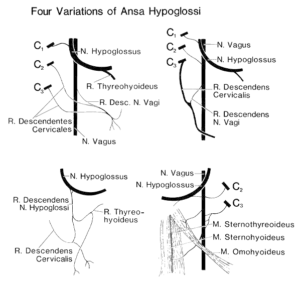
Anatomy Atlases. Immagine ospitata dal sito d’origine. Apri la pagina originale.
Classificazione anatomica
- Sistemi:
- Sistema nervoso
Provenienza del contenuto
Adattamento
Adattamento di «Cervical Plexus — Illustrated Encyclopedia of Human Anatomic Variation: Opus I: Muscular System» di Ronald A. Bergman, Adel K. Afifi, Ryosuke Miyauchi. Fonte: https://www.anatomyatlases.org/AnatomicVariants/NervousSystem/Text/CervicalPlexus.shtml Opera originale distribuita con licenza CC BY-NC-SA 4.0 (https://creativecommons.org/licenses/by-nc-sa/4.0/deed.it). Adattamento a cura di Redazione AIVA. Modifiche rispetto all’originale: Testo originale inglese riprodotto integralmente e riorganizzato nel formato scheda del portale (sintesi, testo, riferimenti), senza modificarne le affermazioni. La traduzione italiana non è ancora stata eseguita, e la pagina lo dichiara. Le figure non sono state copiate: sono collegate alla pagina d’origine. Questa pubblicazione non è approvata né avallata dagli autori dell’opera originale.
Licenza
Questo contenuto è distribuito con licenza CC BY-NC-SA 4.0. Licenze e attribuzioni