Voce enciclopedica
Vertebre cervicali
Abstract
Le vertebre cervicali presentano notevole variabilità dei processi spinosi, dei forami trasversari e dei rapporti fra atlante, epistrofeo e osso occipitale, con possibili fusioni, assimilazione dell’atlante e os odontoideum. Il processo costale della settima vertebra può essere sostituito da una costa cervicale di forma variabile.
Le vertebre cervicali mostrano una variabilità considerevole nei loro processi spinosi. Di regola, nella razza bianca, la seconda, la terza, la quarta e la quinta vertebra possiedono spine bifide.
La sesta e la settima manifestano una tendenza a essere bifide con due piccoli tubercoli laterali. La sesta ha occasionalmente una spina bifida, la settima raramente. Occasionalmente tutte le spine cervicali, a eccezione della seconda, sono non bifide.
Nelle «razze di colore» le spine cervicali sono in genere relativamente più corte che nei bianchi e, di regola, sono semplici. L’unica vertebra cervicale che presenta una spina bifida in tutte le razze è l’epistrofeo. Può essere non bifida nella razza nera, ma raramente nella razza bianca.
Possono verificarsi vertebre cervicali a blocco o fuse. In uno studio furono riscontrate tre fusioni fra la seconda e la terza vertebra (3%) e due fra la quinta e la sesta (2%). La vertebra a blocco mostrava fusione dei corpi vertebrali e delle lamine, mentre spine, processi trasversi e peduncoli restavano indipendenti. Le anomalie vertebrali associate comprendono la sacralizzazione incompleta della quinta lombare e ampi iati sacrali. Fra i corpi fusi era visibile un disco intervertebrale ipoplasico, ma non vi era alcuna traccia di uno spazio articolare fra le faccette articolari.
Le lamine delle vertebre cervicali inferiori presentano frequentemente dorsalmente tubercoli distinti dai quali originano fascicoli del muscolo multifido. I tubercoli possono trovarsi sulla quarta e sulla quinta vertebra, ma più frequentemente sulla sesta e sulla settima. I tubercoli anteriori sono sporgenze ossee che si possono osservare, di solito, sulla sesta cervicale nelle proiezioni laterali del rachide cervicale.
Un foramen transversarium, tipico dei forami dei processi trasversi delle vertebre cervicali, è stato riscontrato nella prima lombare, nella quinta lombare e nella prima vertebra sacrale.
Un forame trasversario accessorio, posteriore e più piccolo rispetto al forame primario, può trovarsi nella sesta vertebra, meno frequentemente nelle vertebre adiacenti
Il forame trasversario della settima cervicale è tipicamente piccolo e può essere assente. Raramente trasmette l’arteria vertebrale, ma frequentemente consente il passaggio di una vena.
L’atlante dell’uomo, confrontato con quello di altri animali, è considerato un «osso degenerato».
L’atlante può mostrare un’ossificazione incompleta dell’arco anteriore e posteriore. L’assenza dell’arco anteriore è stata riportata in un adulto. L’arco posteriore può possedere faccette che si articolano con faccette presenti sul margine del forame magno. Anche l’arco anteriore può avere faccette che si articolano con sporgenze dell’osso occipitale.
Si riscontra comunemente il solco per l’arteria vertebrale trasformato in un forame. Sono stati riportati ossicini pari nella membrana atlo-occipitale posteriore, interpretati come vestigia di un proatlante.
Può verificarsi la fusione, parziale o completa, dell’atlante con l’osso occipitale (incidenza di circa l’1%) (cosiddetta assimilazione dell’atlante).
Per quanto riguarda l’epistrofeo, il dente può formare un os odontoideum separato.
Il sulcus arteriae vertebralis dell’arco posteriore può essere scavalcato da un processo osseo proveniente dal processo articolare inferiore, che produce un canale osseo per l’arteria vertebrale (10% dei casi). Un solco (o canale) retrotrasverso può occasionalmente essere presente sulla massa laterale dell’atlante, posteriormente alla radice del suo processo trasverso. In uno studio (Gupta e coll., 1979) su 123 atlanti secchi l’incidenza fu del 44%. Il solco era presente in 31 reperti (25%), il canale in 14 (11,4%) e canale e solco insieme in 9 (7%). In un altro studio (Veleanu e coll., 1977) su atlanti rumeni l’incidenza fu di circa il 68%. Esso accoglie una vena anastomotica.
Il tubercolo posteriore dell’atlante è raramente bifido, ma può occasionalmente presentare due tubercoli.
L’atlante può essere fuso con l’osso occipitale in gradi variabili. L’«assimilazione» dell’atlante può verificarsi in circa lo 0,5-1% dei crani (1/157).
Il dente dell’epistrofeo può (a) svilupparsi come osso separato, l’os odontoideum; (b) originare dall’arco anteriore; (c) articolarsi con un terzo condilo occipitale; oppure (d) essere assente.
L’epistrofeo (epistropheus) può essere fuso con l’atlante oppure con la terza vertebra.
Il forame vertebro-arterioso associato alla sesta vertebra cervicale appare occasionalmente come un’incisura e può essere associato anche a un forame secondario per la vena vertebrale.
La settima vertebra cervicale (vertebra prominens) è responsabile della prominenza palpabile sul dorso del collo nel 70% degli individui; la sesta cervicale è la successiva per frequenza nel formare tale prominenza (20%) e il processo spinoso della prima toracica è il meno frequente (10%).
Il legamento nucale può contenere osso sesamoide.
Il processo trasverso della settima vertebra può essere bifido e occasionalmente il processo costale è sostituito da una costa cervicale. Il corpo della settima porta talvolta bilateralmente, presso il suo margine caudale, una fossetta costale per la testa della prima costa. Quando è presente una fossetta costale vi è di solito una costa cervicale ben sviluppata. Le coste cervicali associate alla settima vertebra compaiono in forme diverse. Possono essere completamente formate oppure semplici ingrandimenti dei processi costali. In casi molto rari vi è un paio completo di coste, che possono articolarsi con lo sterno. Possono essere rudimentali, presentandosi come una banda fibrosa che si unisce alla consueta prima costa toracica o alla cartilagine della prima costa. La costa cervicale può essere libera alla sua estremità ventrale (distale).
Coste cervicali rudimentali sono state osservate in un caso sulla sesta vertebra cervicale, in due casi sulla quarta, ma mai sulla quinta. Le coste cervicali sono tipicamente presenti nei rettili e negli uccelli.
Varietà di diverse vertebre
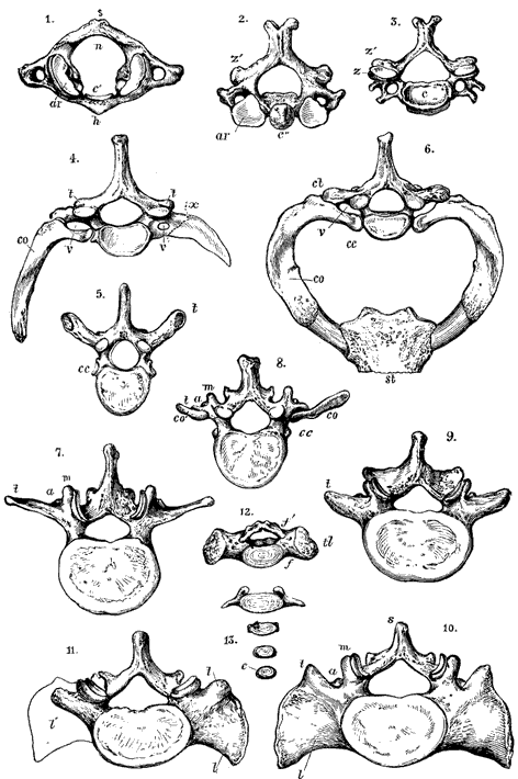
Anatomy Atlases. Immagine ospitata dal sito d’origine. Apri la pagina originale.
Varietà dell’atlante
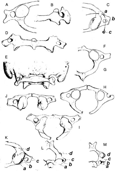
Anatomy Atlases. Immagine ospitata dal sito d’origine. Apri la pagina originale.
Varietà dell’epistrofeo
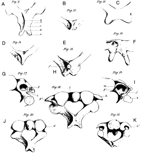
Anatomy Atlases. Immagine ospitata dal sito d’origine. Apri la pagina originale.
Atlante ed epistrofeo
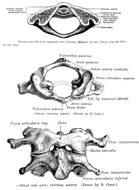
Anatomy Atlases. Immagine ospitata dal sito d’origine. Apri la pagina originale.
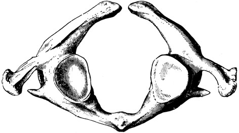
Insolita assenza dell’arco anteriore dell’atlante
Anatomy Atlases. Immagine ospitata dal sito d’origine. Apri la pagina originale.
Coste cervicali

Anatomy Atlases. Immagine ospitata dal sito d’origine. Apri la pagina originale.
Coste cervicali
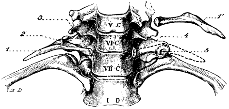
Anatomy Atlases. Immagine ospitata dal sito d’origine. Apri la pagina originale.
Coste cervicali soprannumerarie
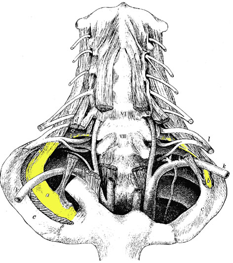
Anatomy Atlases. Immagine ospitata dal sito d’origine. Apri la pagina originale.
Riferimenti dell’opera originale
Elenco riportato come compare nella fonte, senza normalizzazione dei formati bibliografici. Le voci non sono tradotte: un riferimento bibliografico si cita nella lingua in cui è pubblicato.
- Aasar, Y.H. (1937-38) Three cases of fusion of axis with third cervical vertebra. J. Anat. 72:634-635.
- Adson, A.W. (1947) Surgical treatment for symptoms produced by cervical ribs and the scalenus anticus muscle. Surg., Gynecol. Obstet. 85:687-700.
- Adson, A.W. (1851) Cervical ribs: Symptoms, differential diagnosis and indications of sect of the insertion of the scalenus anticus muscle. J. Internat. Coll. Surgeons. 16:546-559.
- Allen, W. (1879) The varieties of the atlas in the human subject, and the homologies of its transverse process. J. Anat. Physiol. 14:18-27.
- Allen, W. (1881) Tertiary occipital condyle. J. Anat. Physiol. 15:60-68.
- Almos, S. (1965) Ungewöhnliche Entwicklungsanomalie des Atlas. Fortschritte Rontgenstrahlen. 102:3 3 1-332.
- Anderson, R.J. (1883) Observations on the diameters of human vertebrae in different regions. J. Anat. Physiol. 17:341-344.
- Barbosa, Sueiro, M.B. (1933) Sur l'osselef odentoidien de l'axis humain. Assoc. Anatomistes Comptes Rendus 28:30-32.
- Bársony, T. and K. Winkler. (1937) Beiträge zur Röntgenologie der Würbelsäule. Röntgenpraxis 9:601-608.
- Baudouin, M. (1915) La deux premières vertièbres cervicales dans l'ossuaire néolithique des cous, a Bazages-en-Pareds (Vendée). Bull. et Mém. de la Soc. d'Anthrop. de Paris 6:5-26.
- Bell, R.H., Swigart, L.L. and B.J. Anson. (1950) The relation of the vertebral artery to the cervical vertebrae. Based upon a study of 200 specimens. Q. Bull. Northwestern University Medical School 24:184-185.
- Bergman, P. (1967) On varieties of the atlas in man. Folia Morphol., Warsaw 26:129-139.
- Bertelsen, S., Mathiesen, F.R. and H.H. Ohlenschlaeger. Vascular complications of cervical rib. Scand. J. Thor. Cardiovasc. Surg. 2:133-139.
- Black, J. (1899) Report of a case of cervical ribs. Proc. Anat. Soc. Gr. Br. Ireland. J. Anat. Physiol. 33:xlvii-1.
- Blair, D.M., Davies, F. and W. McKissock. (1935) The etiology of the vascular symptoms of cervical rib. Brit. J. Surg. 22:406-414.
- Bohlig, H. (1954) Ankylose des Atlanto-Epistropheal-Gelenkes. Fortschr. Röntgenstr. 81:216-218.
- Bohlig, H. and E. Siemon. (1954) Ankylose von Atlas und Epistropheus (Bericht über 1 Fall). Z. Orthop. 85:231-236.
- Borovansky, L. (1928) Les soudures de l'axis et de la troisième vertebrae cervicale chez l'homme. Assoc. Anatomistes Comptes Rendus 23:49-54.
- Bosco, L. (1925) Observations, dérivées de la statistique, sur les manifestations de vertèbres occipitales et sur l'assimilation de l'atlas à l'occipital. Arch. Ital Biol. 75:160.
- Brannon, E.W. (1963) Cervical rib syndrome. An analyse of nineteen cases and twenty four operations. J. Bone Joint Sur. (Am) 45:977-998.
- Brat, L. (1951) Klinische Fehldiagnose durch abnorm langen Griffelfortsatz des Felsenbeins. Fortschr. Röntgenstr. 75:758-759.
- Brocher, J.E.W. (1938) Mehrfach angeborene Fehlbildungen der Wirbelsäule. Fortschr. Röntgenstr. 58:440-447.
- Brocher, J.E.W. (1947) Le processus paracondyloideus s. apophyse paramastoide (dans un cas de dysostose mandibulo faciale unilatérale) Radiol. Clin. 16:393-396.
- Brown, C.E. (1941) Complete absence of the posterior arch of the atlas. Anat. Rec. 81:499-503.
- Buetti, C. (1953) Zur Röntgendiagnostik seltener Fehlbildungen der Halswirbelsäule. Radiol. Clin. 22:141-161.
- Bystrow, A. (1931) Assimilation des Atlas und Manifestation des Proatlas. Z. Anat. Entwickungsgesch. 95:210-242.
- Calori, L. (1896) Sur une union synostotique et une incorporation partielle de l'atlas avec l'os occipital, et sur deux autres anomalies correlatives. Arch. Ital. Biol. 25:152-153.
- Cave, A.J.E. (1925) Fusion of carpal elements. J. Anat. 60:460-461.
- Cave, A.J.E. (1933-34) Anatomical notes on the occipito-atlanto-axial articulations. J. Anat 68:416-423.
- Cave, A.J.E. (1933-34) Cervical intercostal articulation. J. Anat. 68:521-524.
- Cave, A.J.E. (1936-37) A further example of cervical intercostal articulation. J. Anat. 71:415-416.
- Cave, A.J.E. (1941-42) The earliest English example of bilateral cervical rib. Brit. J. Surg. 29:47-51.
- Church, A. (1919) The neurology of cervical rib. JAMA 73:1-4.
- Collins, C.U. (193 1) Cervical ribs. Am. J. Surgery 14:449-451.
- Dale, W.A. (1982) Thoracic outlet compression syndrome. Critique 1982. Arch. Surg. 117:1437- 1445.
- DiDio, L.J.A. (1967) Radiological study of the anatomical variations of the vertebrae prominens in man. Anat. Anz. 120:210-218.
- Diethelm, L. (1943) Zur Kenntnis der Entwicklungsgeschichte der Wirbelsäule und der Wirbelkörperfehlbildungen. Fortschr. Röntgenstr. 69:143-150.
- Donald, J.M. and B.F. Morton. (1940) The scalenus anticus syndrome with and without cervical rib. Ann. Surg. 111:709-723.
- Dubreuil-Chambardel, L. (1907) Variations sexuelles de Patlas. Bull. et Mém. de la Soc. d'Anthrop. de Paris 8:399-404.
- Dubreuil-Chambardel, -. et -. Bonnin. (19 2 1) Colonne cervicale a huit segments vertbraux. Bull. et Mém. de la Soc. d'Anthrop. de Paris 2:29-36.
- Dwight, T. (1887) Account of two spines with cervical ribs, one of which has a vertebra suppressed, and absence of the anterior arch of the atlas. J. Anat. Physiol. 21:539-550.
- Eden, K.C. (1939) The vascular complications of cervical ribs and first thoracic rib abnormalities. Brit. J. Surg. 27:111-139.
- Eggeling, H. von. (1922) Die Gabelung der Halswirbeldornen und ihre Ursache. Anat. Anz. 55:33-94.
- Elliot Smith, G. (1907) On a case of fusion of the atlas and axis. Anat. Anz. 31:166-168.
- Ferrari, G. (1913) Sur la soudure de l'os occipitis avec l'atlas. Arch. Ital. Biol. 59:472.
- Flecker, H. (1942) Time of appearance and fusion of ossification centers as observed by roentgenographic methods. Am. J. Roentgenol. 47:97-159.
- Francis, C.C. (1955) Variations in the articular facets of the cervical vertebrae. Anat. Rec. 122:589-602.
- Francis, C.C. (1955) Dimensions of the cervical vertebrae. Anat. Rec. 122:603-609.
- Frank, J. (1914-15) Ein Fall von Halsrippe mit abnormen Nervenverlauf. Anat. Anz. 47:218-225.
- Friedlowsky, A. (1869) über die sogannater accessorischen Gelenkshöcker an der Pars basiliaris ossis occipitis und einige Formen von ungewöhnhcher Gelenksverbindung zwischen den Zahnfortsatz des Epistropheus und dem Hinterhauptknochen. Wiener Akad. Sitzungberichte. Math.-Natur. Kl. 60:319- 342.
- Fullenlowe, T.M. (1954) Congenital absence of the odentoid process. Report of a case. Radiology 63:72-73.
- Funke, E. (1898) über einen Processus odentoideus atlantis hominis. Anat. Anz. 14:385-390.
- Geipel, P. (1930) Zur Kenntnis der Spina bifida am Atlas. Fortschr. Röntgenstr. 42:583-589.
- Geipel, P. (1932) Zur Kenntnis der Spaltbildung des Atlas und Epistropheus. Fortschr. Röntgenstr. 46:373-402.
- Gershon-Cohen, J. and R.F. Delbridge. (1945) Pseudoarthrosis, synchrondrosis and other anomalies of the first ribs. Am. J. Roentgenol. 53:49-54.
- Giacomini, -. (1887) De l'existence de l'os odontoïde chez l'homme. Arch. Ital. Biol. 8:40-48.
- Giannestras, N.J., Mayfield, F.H., Provencio, F.P. and J. Mauer. (1955) Congenital absence of the odontoid process. Case report. J. Bone Joint Surg. (Am) 37:839.
- Gilliatt, R.W.;'Willison, R.G., Dietz, V. and I.R. Williams. (1978) Peripheral nerve conduction in patients with cervical rib and band. Ann. Neurol. 4:124-129.
- Gillman, E.L. (1959) Congenital absence of the odentoid process of the axis. J. Bone Joint Surg. (Am) 41:345-348.
- Gladstone, R.J. and C.P.G. Wakeley. (1931-32) Cervical ribs and rudimentary first thoracic ribs considered from clinical and etiological standpoints. J. Anat. 66:334-370.
- Gockel, H.P. (1958) über eine Form von Atlasfehlbildung. Fortschr. Röntgenstr. 88:485-487.
- Goodhart, S.P. (1909) Cervical rib and its relation to the neurpathies. Am. J. Med Sci. 138:666-683.
- Grashey, R. (1935) Fehlerquellen, Varietäten Rippenanomalie: artikulierende Halsrippe. a) Hornförmiger Auswuchs am Os navic, manus. b) Abbruch der oberen Gelenkante des Os navic. manus; Lücke im Karpus. Rontgenpraxis 7:209, 275.
- Green, H.L.H.H. (1930) An unusual case of atlanto-occipital fusion. J. Anat. 65:140-144.
- Gruber, W. (1851) Über den gesamten Apparat der Bänder zwischen dem Hinterhauptsbein und einem neuentdeckten Appendix superior des Ligamentum cruciatum inbesondere. Arch. Anat. Physiol. Wissen. Med. 1851:291-315.
- Gruber, W. (1876) Über ein Gelenk zwischen den vorderen Querfortsätzen der rechten Seite des V. und VII. Halswirbels. Arch. Pathol. Anat. Physiol. Klin. Med. 67:327-330.
- Gruber, W. (1877) Verwachsung zweier Rippen durch eine congenitale Knochenplatte zwischen deren Hälsen. Arch. Pathol. Anat. Physiol. Klin. Med. 70:139-140.
- Gupta, S.C., Gupta, C.D., Arora, A.K. and B.B. Maheshwari. The retrotransverse groove (canal) in the Indian atlas vertebrae. Anat. Anz. 145:514-516.
- Hadley, L.A. (1948) Atlanto-occipital fusion, ossiculum terminale and occipital vertebra as related to basilar impression with neurological symptom. Am. J. Roentgenol. 59:511-524.
- Hertslet, L.E. and A. Keith. (1896) Comparsion of anomalous parts of two subjects, one with a cervical rib, the other with a rudimentary first rib. J. Anat. Physiol. 30:562-567.
- Hill, R.M. (1939) Vascular anomalies of the upper limb associated with cervical rib: Report of a case and review of the literature. Brit. J. Surg. 27:100-110.
- Hohl, K. (1959) Das Os odontoideum (partielle Densplasie). Fortschr. Röntgenstr. 91:518-521.
- Hunter, R.H. (1924) An abnormal atlas. J. Anat. 58:140-141.
- Ingelmark, B.E. (1947) Über das craniovertebral Grenzgebeit beim Menschen. Acta Anat. [Suppl.] 6:1-116.
- Ivie, J. McK. (1946) Congenital absence of the odontoid process. Report of a case. Radiology 46:268-269.
- Jacquemet, -. (1850) Anomalie de l'articulation axoïde-occipitale. Bulletins et Mem. de la Société Anatomique de Paris XXV(2):49.
- Jain, S.P. (1956) A rare and unusual variety of atlanto-occipital fusion. J. Anat. Soc. India 5:72-73.
- Janker, R. (1935) Eine anatomische Variante am Querforsatz des Atlas. Röntgenpraxis 7:399.
- Karlen, A. (1962) Congenital hypoplasia of the odontoid process. J. Bone Joint Surg. (Am) 44:567-570.
- Keating, D.R. and J.R. Amberg. (1954) A source of potential error in the roentgen diagnosis of cervical ribs. Radiology 62:688-694.
- Keen, W.W. (1907) The symptomology, diagnosis, and surgical treatment of cervical ribs. Am. J. of the Med. Sci. 133:173-218.
- Keller, H.L. (1960) Eine seltene Form der Manifestation des Okizipitalwirbels. Fortschr. Röntgenstr. 93:370-372.
- Kendrick, G.S. and N.L. Biggs. (1963) Incidence of the pontiuculus posticus of the first cervical vertebra between the ages of six to seventeen. Anat. Rec. 145:449-453.
- Kippel, -. (1912) Un cas d'absence des vertèbres cervicales cage thoradque remontant jusqu'a la base du crâne. Bull. et Mém. de la Soc. d'Anthrop. de Paris. 3:101-102.
- Kolte, D.T. and V.R. Mysorekar. (1963) Cervico-occipital fusions. J. Anat. Soc. India 12:36-39.
- Kostanecki, K. von. (1891) Beiträge zur Kenntnis der Missbildungen in der Kopf- und Halsgegend. Arch. Pathol. Anat. Physiol. Klin. Med. 123:401-428.
- Krisch, H. (1918) Rudimentäre Halsrippen beiderseits und Otosklerose. Dtsch. Med. Wochenschr. 44:670.
- Lanier, R.R. jr. (1939) An anomalous cervico-occipital skeleton in man. Anat. Rec. 73:189-201.
- Lapayowker, M.S. (1960) An unusual variant of the cervical spine. Am. J. Roentgenol. 83:656-659.
- Law, A.A. (1920) Adventitious ligaments simulating cervical ribs. Am. Surg. 72:497-499.
- Ledenyi, J. (1939) Ankylosis articuli atlantooccipitalis sinistra. Anat. Anz. 88:503-505.
- LeDouble, A.F. (1911) Cotes cervicales chez Phomme. Bull. et Mém. de la Soc. d'Anthrop. de Paris. 2:501-533.
- LeDouble, A.F. (1912) Soudure chez Phomme de Patlas a la base du crâne. Bull. et Mém. de la Soc. d'Anthrop. de Paris. 3:20-35.
- Lucas, M.F. (1915) (1) Two cases of cervical ribs. (2) An anomalous arrangement of the vagi. J. Anat. Physiol. 69:336-342.
- Macalister, A. (1893) Notes on the development and variations of the atlas. J. Anat. Physiol. 27:519-542.
- Macalister, A. (1894) The development and variations of the second cervical vertebrae. J. Anat. Physiol. 28:257-268.
- McRae, D.L. and A.S. Barnum. (1953) Occipitalization of atlas. Am. J. Roentgenol. 70:23-46.
- Mex, W. (1959) Ungewöhnliche Form zweier Halsrippen. Monatschr. Unfallheilk, Versicherungsmed. 62:307-310.
- Misra, B.D. (1954) A case of atlanto-occipital fusion. J. Anat. Soc. India 3:45-46.
- Miyakawa, G. (1952) Congenital absence of the odentoid process. A case report. J. Bone Joint Surg. (Am) 34:676-677.
- Monteiro, H. (1933) Fréquence de l'occipitalisation de l'atlas chez les Portugais. Assoc. Anatomistes Comptes Rendus 28:465-470.
- Nayak, U. V. (1931) Anatomical notes. A case of abnormal atlas and axis vertebra. J. Anat. 65:399-400.
- Noback, C.R. and G.G. Robertson. (1951) Sequences of appearance of ossification centers in the human skeleton during the first five prenatal months. Am. J. Anat. 89:1-28.
- Okamoto, K. (1924) über das Foramen transversarium und die Eigentümlichkeit des Querfortsatzes am 7 Halswirbel. Anat. Anz. 58:401-430.
- Osler, W. (1910) Certain vasomotor, sensory and muscular phenomena associated with cervical ribs. Am. J. of the Med. Sci. 139:469-472.
- Overton, L.M. and J.W. Grossman. (1952) Anatomical variations in the articulation between the second and third cervical vertebrae. J. Bone Joint Surg. (Am) 34:155-161.
- Pancoast, H.K. (1901-02) Cervical rib. University Penn. Med. Bull. 14:394-398.
- Pate, J.R. (1936-37) An unusual occipito-atloid articulation. J. Anat. 71:128-129.
- Patterson, R.H. (1940) Cervical ribs and the scalenus muscle syndrome. Ann. Surg. 111:531-545.
- Phoebus, P. (1835) Zur pathologischen anatomie. Novi Acta Physico-Medica Academiae Caesareae Leopoldino-Carolinae Naturae Curiosorum 17(2):665-674.
- Pitzorno, M. (1899) L'épistrophée. Arch. Ital. Biol. 31:179.
- Pitzorno, M. (1899) Sur quelques novelles particularités da l'atlas et sur la coexistence de ses variétés. Arch. Ital. Biol. 31:179.
- Purves, R.K. and P.H. Wedin. (1950) Familial incidence of cervical ribs. J. Thoracic Surg. 19:952-956.
- Rau, R.K. and D. Sivasubrahmanian. (1933) Anomalous atlantoocciput. J. Anat. 67:622-623.
- Reinhardt, K. (1956) Eine ungewöhnliche Anomalie an den Dornfortsätzen des 5. 6. und 7. Halswirbels. Fortschr. Röntgenstrahlen 85:253-255.
- Renander, A. (1929) Anomalies roentgenologically observed of the craniovertebral region. Acta Radiol. 10:502-513.
- Riches, E.W. (1928-29) The anatomy of cervical rib: With a case report. Brit. J. Surg. 16:235-238.
- Roberts, S.M. (1933) Congenital absence of the odentoid process resulting in dislocation of the atlas on the axis. J. Bone Joint Surg. (Am) 15:988-989.
- Roche, M.B. and G.G. Rowe. (1952) The incidence of separate neural arch and coincident bone variations. A summary. J. Bone Joint Surg. (Am) 34:491-494.
- Romanus, T. and A. Tori. (1964) A variation of the atlas: Roentgenalogic incidence of a bridge over the groove on the atlas for the vertebral artery. Acta Radiol. Diag. 2:289-297.
- Ross, Sir J.P. (1959) The vascular complications of cervical rib. Ann. Surg. 150:340-345.
- Rousseaux, R., Kissel, P., Beau, A., Midon, J. and G. Arnould. (1950) Syntose occipito-atloïdiene; rétécissement du trou occipital et compression bulbo-médullaire par proéminences osseuses, vestiges des masses latérales de l'atlas. Rev. Neurol. 82:389-394.
- Saunders, W.W. (1943) Basilar impression: The position of the normal odontoid. Radiology 41:589-590.
- Scannell, R.C. (1945) Congenital absence of the odentoid process. A case report. J. Bone Joint Surg. 27:714-715.
- Schmidt, H. and E. Fischer. (1960) über partielle einseitige Synostosen zwischen Atlas und Axis. Fortschr. Röntgenstr. 92:380-384.
- Schultz, E.H., Jr., Levy, R.W. and P.E. Russo. (1956) Agenesis of the odontoid process. Radiology 67:102-105.
- Selby, S., Garn, S.M. and V. Kanareff. (1955) The incidence and familial nature of a bony bridge on the first cervical vertebrae. Am. J. Phys. Anthroplol. 13:129-141.
- Shehata, R. (1964) Occipitalisation of the atlas. J. Anat. Soc. India 13:97-99.
- Shore, L.R. (1931) A report on the spinous processes of the cervical vertebrae in the native races of South Africa. J. Anat. 65:483-505.
- Shore, L.R. (1933) A report on the spinous processes of the cervical vertebrae in a series of Egyptian skeletons. J. Anat. 67:422-442.
- Short, D.W. (1975) The subclavian artery in 16 patients with complete cervical ribs. J. Cardiovasc. Surg. 16:135-141.
- Singh, S. (1965) Variations of the superior articular facets of atlas vertebrae. J. Anat. 99:565-571.
- Smith, E.G. (1907) On a case of fusion of the atlas and axis. Anat Anz. 31:166-168.
- Stanczyk, J.L. (1979) Canal of vertebral artery of atlas. Folia Morphol., Warsaw 38:353-357.
- Stein, M. (1911) über einen Fall von beiderseitigen Halsrippen. Anat. Anz. 38:650-664.
- Stevenson, P.H. (1924) Age order of epiphyseal union in man. Am. J. Phys. Anthropol. 7:53-93.
- Stiefel, D.M. (1950) Congenital absence of the odontoid process. Report of a case. J. Bone Joint Surg. (Am) 32:946-947.
- Swjetschnikow, -. (1906) über die Assimilation des Atlas und die Manifestation des Occipitalwirbels beim Menschen. Arch. Anat. Physiol. Wissen. Med. 1906:155-194.
- Telford, E.D. and J.S.B. Stopford. (193 0-3 1) The vascular complications of cervical rib. Brit. J. Surg. 18:557-564.
- Teplick, J.G., Steinberg, S. and B.P. Adelman. (1960) Congenital absence of the odontoid process. Am. J. Roentgenology 83:653-655.
- Todd, T.W. (1897) "Cervical rib": Factors controlling its presence and its size. Its bearing on the morphology and development of the shoulder (with four cases). J. Anat. Physiol. 31:288-302.
- Todd, T.W. (1911) The relations of the thoracic operculum considered in reference to the anatomy of cervical ribs of surgical importance. J. Anat. Physiol. 45:293-304.
- Todd, T.W. (1912) Costal anomalies of the thoracic inlet, their interpretation and significance. Anat. Anz. 41:257-271.
- Todd, T.W. (1912) "Cervical" rib: Factors controlling its presence and its size. Its bearing on the morphology and development of the shoulder. J. Anat. 46:244-288.
- Todd, T.W. (1914) L'apophyse transverse de la 7 e vertèbres cervicale et ses rapports morphologiques avec les apophyses transverses des vertèbres sus-jacente. Bull. et Mém. de la Soc. d'Anthrop. de Paris 5:282-294.
- Todd, T.W. et j. D'Errico, Jr. (1926) The odontoid ossicle of the second cervical vertebra. Ann. Surg. 83:20-31.
- Tramontano-Guerritore, G. (1927-28) Die Atlanto-occipital-union. Anat. Anz. 64:173-184.
- Trolard, P. (1892) Note sur la présence d'un petit arc osseux dans l'épaisseur du ligament atloido-occipital postérieur. Soc. Biol. Comptes Rendus Hebdomadaires des Séances et Mémoires 44:226.
- Turner, W. (1870) Supernumerary cervical ribs. J. Anat. Physiol. 4:130-139.
- Veleanu, C., Barzu, S., Panescu, S. and C. Udroiu. (1977) The retrotransverse groove or canal of the atlas and its significance. Acta Anat. (Basel) 97:400-407.
- Viehweger, G. (1955) Der Processus paratransversarius, eine Varietät am Atlas. Fortschr. Röntgenstr. 83:411-412.
- Volkman, J. (1934) über den Processus styloideus der wirbel. Zentralbl. Chir. 61:1340-1342.
- Walshe, F.M.R., Jackson, H. and R. Wyburn-Mason. (1944) On some pressure effects associated with cervical and rudimentary and "normal" first ribs, and the factors entering into their causation. Brain 67:141.
- Weiler, H.G. (1942) Congenital absence of odontoid process of the axis with atlanto-axial dislocation. J. Bone Joint Surg. (Am.) 24:161-165.
- Wells, T.R., Jacobs, R.A., Senac, M.O. and B.H. Landing. (1990) Incidence of short trachea in patients with myelomeningocele. Pediatric Neurol. 6(2): 109-111.
- Went, H. (1958) Zum klinischen Bild der Atlasassimilation. Fortschr. Röntgenstr. 89:213-219.
- Whitney, C. (1926) Asymmetry of vertebral articular processes and facets. Am. J. Phys. Anthropol. 9:451-455.
- Wollin, D.G. (1963) The os odontoideum. Separate odontoidal process. J. Bone Joint Surg. (Am.) 45:1459-1471.
- Wood-Jones, F. (1937-38) The cervical vertebrae of the Australian native. J. Anat. 72:411-415.
- Zaborowski, Z. (1975) The sagittal foramen of the atlas. Folia Morphol., Warsaw 34:375-381.
Classificazione anatomica
- Sistemi:
- Sistema scheletrico
Provenienza del contenuto
Traduzione
Traduzione italiana di «Cervical Vertebrae — Illustrated Encyclopedia of Human Anatomic Variation: Opus I: Muscular System» di Ronald A. Bergman, Adel K. Afifi, Ryosuke Miyauchi. Fonte: https://www.anatomyatlases.org/AnatomicVariants/SkeletalSystem/Text/CervicalVertebrae.shtml Opera originale distribuita con licenza CC BY-NC-SA 4.0 (https://creativecommons.org/licenses/by-nc-sa/4.0/deed.it). Traduzione a cura di Redazione AIVA. Modifiche rispetto all’originale: Traduzione italiana del testo originale inglese, che resta consultabile nella versione inglese di questa pagina. Il contenuto è stato riorganizzato nel formato scheda del portale (sintesi, testo, riferimenti) senza modificarne le affermazioni. Le figure non sono state copiate: sono collegate alla pagina d’origine. Questa pubblicazione non è approvata né avallata dagli autori dell’opera originale.
Licenza
Questo contenuto è distribuito con licenza CC BY-NC-SA 4.0. Licenze e attribuzioni
