Voce enciclopedica
Facial (CN VII) Nerve
Abstract
A pattern of variation has been established for the facial nerve (see Illustrations). However, the clear separation of the nerve into five main terminal branches (temporal, zygomatic, buccal, mandibular, and cervical) may be difficult or impossible. The posterior auricular and occipital branches may arise before their exit from the stylomastoid foramen. Branches from the facial nerve have been observed to replace those from the glossopharyngeal (CN IX) in supplying the root of the tongue and palate.
A pattern of variation has been established for the facial nerve (see Illustrations). However, the clear separation of the nerve into five main terminal branches (temporal, zygomatic, buccal, mandibular, and cervical) may be difficult or impossible.
The posterior auricular and occipital branches may arise before their exit from the stylomastoid foramen.
Branches from the facial nerve have been observed to replace those from the glossopharyngeal (CN IX) in supplying the root of the tongue and palate.
The nerve to the digastric muscle may also supply the styloglossus and, in part, the sternocleidomastoid.
The facial nerve may furnish branches to CN X (Vagus) and CN XI (spinal accessory) nerves.
In a study of 78 preparations, the chorda tympani originated from the facial nerve within the facial canal in 74, and extracranially in four.
The chorda tympani may run independently of the lingual nerve after its exit from the iter chordae anterius and the petrotympanic fissure.
It has been observed attached to the inferior alveolar nerve until the latter entered the alveolar canal; the chorda tympani then supplied the salivary glands and joined the lingual to supply the tongue.
The facial nerve may have an unusual course through the temporal bone; it may be exposed in the middle ear by lacking a bony cover, and it may be divided into several parts and be in separate canals. Unusual muscle fibers may be found in the facial canal along with the facial nerve.
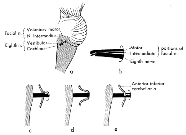
Image 24A Image 24B
Anatomy Atlases. Immagine ospitata dal sito d’origine. Apri la pagina originale.
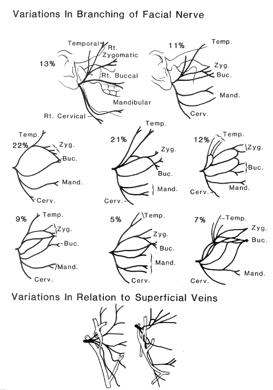
Image 24A Image 24B
Anatomy Atlases. Immagine ospitata dal sito d’origine. Apri la pagina originale.
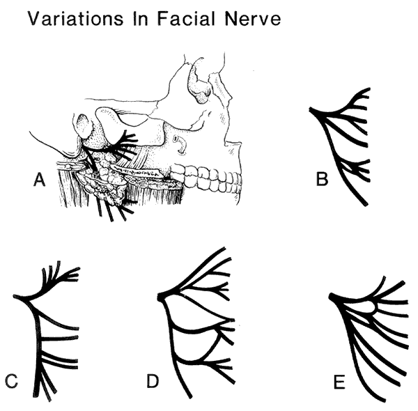
Image 24A Image 24B
Anatomy Atlases. Immagine ospitata dal sito d’origine. Apri la pagina originale.
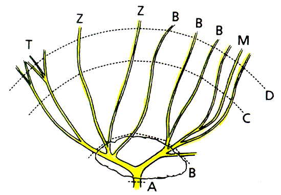
Image 24A Image 24B
Anatomy Atlases. Immagine ospitata dal sito d’origine. Apri la pagina originale.
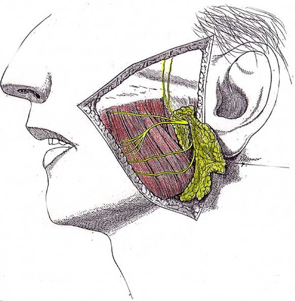
Image 24A Image 24B
Anatomy Atlases. Immagine ospitata dal sito d’origine. Apri la pagina originale.
Riferimenti dell’opera originale
Elenco riportato come compare nella fonte, senza normalizzazione dei formati bibliografici.
- Adams, S.B., Wheeler, J.F.G. and F.H. Edgeworth. (1929) On the innervation of the platysma and mandibulo-auricularis. J. Anat. 63:242-252.
- Anson, B.J., Ed. (1966) Morris' Human Anatomy, 12th ed. The Blakiston Division, McGraw-Hill Book Co., New York.
- Bailey, H. (1941) The treatment of tumors of the parotid gland with special reference to total parotidectomy. Br. J. Surg. 28:337-346.
- Banfai, P. (1976) Die angeandte klinische Anatomie des Nervus facilis. I. Kernsystem, supranucleare Verbingung, Peripherie. (Applied Anatomy of the Facial Nerve. I. Nuclei, Supranuclear connectiosn and peripherial nerve.) HNO 24(8):253-264.
- Banfai, P. (1976) Die angeandte klinische Anatomie des Nervus facilis. II Die Anastomosen. (Applied Anatomy of the Facial Nerve. II. Anastomoses.) HNO 24(9):289-294.
- Cannieu, -. (A.) (1885) Remarques sur le nerf intermediaire de Wrisberg. Comp. Rend. Acad Sci., Paris 120:880-882.
- Cruveilhier, J (1851) Traite d'Anatomie Descriptive, 3rd ed. G. Doin & Cie., Paris.
- Cushing, H. (1904) The sensory distribution of the fifth cranial nerve. Johns Hopkins Hospital Bulletin 15:213-232.
- Dargent, M. et P.E. Duroux. (1946) Donnés anatomiques concernant la morphologie et certains rapports du facial intra-parotiden. La Presse Médicale No. 37, 1946:523-524.
- Davis, R.A., Anson, B.J., Budinger, J.M. and LeR. E. Kurth. (1956) Surgical anatomy of the facial nerve and parotid gland based upon a study of 350 cervicofacial halves. Surg. Gynecol. Obstet. 102:385-412.
- Grégoire, R. (1912) Le nerf et la parotide. J. de l'Anatomie et de la Physiol. Norm. Pathol. de l'Homme et des Animaux 48::437-447.
- Gruber, W. (1872) Ungewöhnliche Lage des Nervus facialis in der Parotis zu den Gefässen. Arch. f. Pathologische Anatomie u. Physiol. u. f. Klinische Medicine 45(2):190.
- Gruber, W. (1872) Ungewöhnliche Lage des Nervus facialis in der Parotis zu den Gefässen. Arch. Pathol. Anat. Physiol. Klin. Med. 54:190.
- Henle, J. (1868) Handbuch der Systematischen Anatomie des Menschen. von Friedrich Vieweg und Sohn, Braunschweig.
- Hurford, F.R. (1946) The surgical anatomy of the parotid gland. Brit. J. Surg. 34:186-187.
- Klein, M. (1946) Surgical anatomy of the facial nerve. Br. J. Ophthalmology 30:668-675.
- Kulczynski, B. and W. Wozniak. (1978) Variation of the origin and course of the chorda Tympani. Folia Morphol., Warsaw 37:237-241.
- Latarjet, A. (1948) Testut's Traité d'Anatomie Humaine, 9th ed. G. Doin & Cie., Paris.
- McCormack, L.J., Cauldwell, E.W. and B.J. Anson. (1945) The surgical anatomy of facial nerve with special reference to the parotid gland. Surg. Gynecol. Obstet. 80:620-630.
- McKenzie, J. (1948) The parotid gland in relation to the facial nerve. J. Anat. 82:183-196.
- Minnigerode, B. (1965) Intratympanic Aurzweigung der Chorda Tympani. Ein Seltene Anatomische Verlaufsvariante. Acta Otolaryngol. 59:20-22.
- Oliver, G., Aboulker, P. and Eyries. (1953) Les différents types de nerf facial intra-parotidien. Assoc. Anatomistes Comptes Rendus 40:859-869.
- Patey, D.H. and I. Ranger. (1957) Some points in the surgical anatomy of the parotid gland. Br. J. Surg. 45:250-258.
- Rudolph, R. (1990) Depth of the facial nerve in face lift dissections. Plast. Reconstr. Surg. 85(4):537-544.
- Saltzstein, H.C. (1936) Total removal of parotid gland with preservation of facial nerve. Ann. Surg. 103:635-638.
Classificazione anatomica
- Sistemi:
- Sistema nervoso
Provenienza del contenuto
Adattamento
Adattamento di «Facial (CN VII) Nerve — Illustrated Encyclopedia of Human Anatomic Variation: Opus I: Muscular System» di Ronald A. Bergman, Adel K. Afifi, Ryosuke Miyauchi. Fonte: https://www.anatomyatlases.org/AnatomicVariants/NervousSystem/Text/FacialNerve.shtml Opera originale distribuita con licenza CC BY-NC-SA 4.0 (https://creativecommons.org/licenses/by-nc-sa/4.0/deed.it). Adattamento a cura di Redazione AIVA. Modifiche rispetto all’originale: Testo originale inglese riprodotto integralmente e riorganizzato nel formato scheda del portale (sintesi, testo, riferimenti), senza modificarne le affermazioni. La traduzione italiana non è ancora stata eseguita, e la pagina lo dichiara. Le figure non sono state copiate: sono collegate alla pagina d’origine. Questa pubblicazione non è approvata né avallata dagli autori dell’opera originale.
Licenza
Questo contenuto è distribuito con licenza CC BY-NC-SA 4.0. Licenze e attribuzioni