Voce enciclopedica
Internal Pudendal Artery
Abstract
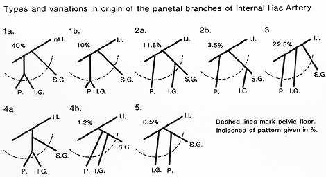
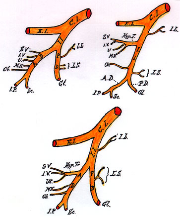
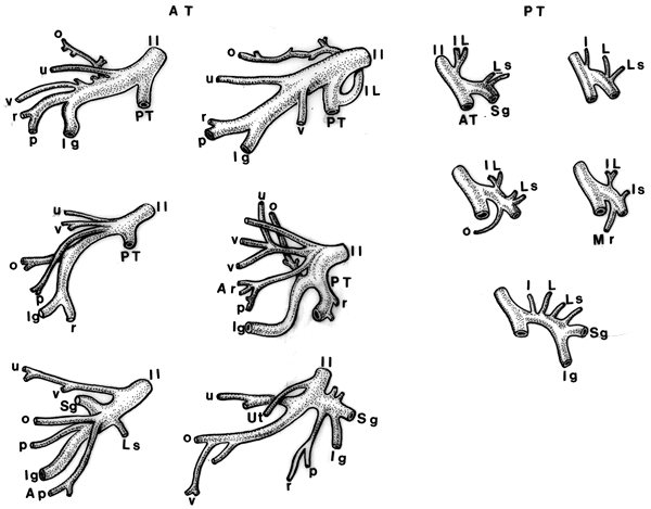
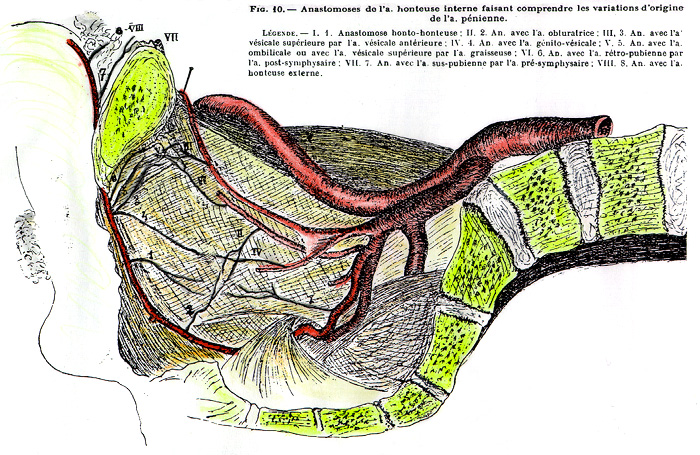
The internal pudendal artery may arise in common with the obturator or the umbilical. It may end as a perineal artery, with the artery to the penis arising from the internal iliac. In some cases, it splits into two vessels, one of which remains in the pelvis to supply pelvic viscera. The internal pudendal is necessarily, always a branch of the anterior division of the internal iliac artery. The dorsalis penis artery may arise from profunda femoris or femoral artery. The inferior rectal artery is typically a branch of the internal pudendal artery.
The internal pudendal artery may arise in common with the obturator or the umbilical.
It may end as a perineal artery, with the artery to the penis arising from the internal iliac.
In some cases, it splits into two vessels, one of which remains in the pelvis to supply pelvic viscera.
The internal pudendal is necessarily, always a branch of the anterior division of the internal iliac artery.
The dorsalis penis artery may arise from profunda femoris or femoral artery.
The inferior rectal artery is typically a branch of the internal pudendal artery.
The Number of Rectal Arteries in Man   Authors   Levi Dubreuil-Chambardel One inferior rectal artery 44% 50% Two inferior rectal arteries 52% 41% Three inferior rectal arteries 3% 5% Four inferior rectal arteries 1% 2% Five inferior rectal arteries 1% 2%
External Pudendal Superficial
Anatomy Atlases. Immagine ospitata dal sito d’origine. Apri la pagina originale.
External Pudendal Superficial
Anatomy Atlases. Immagine ospitata dal sito d’origine. Apri la pagina originale.
External Pudendal Superficial
Anatomy Atlases. Immagine ospitata dal sito d’origine. Apri la pagina originale.
External Pudendal Superficial
Anatomy Atlases. Immagine ospitata dal sito d’origine. Apri la pagina originale.
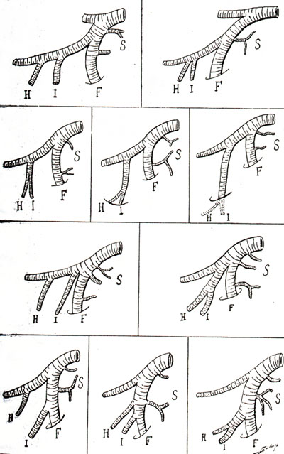
External Pudendal Superficial
Anatomy Atlases. Immagine ospitata dal sito d’origine. Apri la pagina originale.
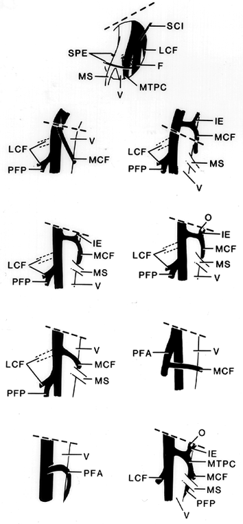
Rectal Inferior
Anatomy Atlases. Immagine ospitata dal sito d’origine. Apri la pagina originale.
See Image 413
Riferimenti dell’opera originale
Elenco riportato come compare nella fonte, senza normalizzazione dei formati bibliografici.
- In addition to those listed for the inferior vesical artery , see also
- Dschau, F. (1936-37) Eine bisher unbekannte Varietät der Arteria obturatoria und pudenda accessoria mit einer Varietät der A. circumflexa femoris medialis. Anat. Anz. 83:25-29.
- Flesch, M. (1876) Varietaten-Beobachtungen aus dem Praparirsaal zu Wurzburg in der Zeit vom 1. Februar 1874 bis 1. April 1875. Verhandlungen der Physikalisch-Medicinische Gesellschaft im Wurzburg 10:25-62.
- Parsons, F.G. and A. Keith. (1897) Sixth annual report of the Committee of Collective investigation of the Anatomical Society of Great Britain and Ireland. J. Anat. Physiol., London 31:31-44.
- Ramsay, A. (1813) Account of unusual conformation of some muscles and vessels. Edinburgh Medical and Surgical Journal 8:281-283.
- Walsham, W.J. (1880) An accessory pudic artery crossing the middle line of the body immediately above and in front of the prostate. St Bartholomews Hospital Reports 16:92-93.
- Winslow, R. (1883) A study of the malformations, variations, and anomalies of the circulatory apparatus in man. Annals of Anatomy and Surgery 7:148-149.
Classificazione anatomica
- Sistemi:
- Sistema cardiovascolare
Provenienza del contenuto
Adattamento
Adattamento di «Internal Pudendal Artery — Illustrated Encyclopedia of Human Anatomic Variation: Opus I: Muscular System» di Ronald A. Bergman, Adel K. Afifi, Ryosuke Miyauchi. Fonte: https://www.anatomyatlases.org/AnatomicVariants/Cardiovascular/Text/Arteries/PudendalInternal.shtml Opera originale distribuita con licenza CC BY-NC-SA 4.0 (https://creativecommons.org/licenses/by-nc-sa/4.0/deed.it). Adattamento a cura di Redazione AIVA. Modifiche rispetto all’originale: Testo originale inglese riprodotto integralmente e riorganizzato nel formato scheda del portale (sintesi, testo, riferimenti), senza modificarne le affermazioni. La traduzione italiana non è ancora stata eseguita, e la pagina lo dichiara. Le figure non sono state copiate: sono collegate alla pagina d’origine. Questa pubblicazione non è approvata né avallata dagli autori dell’opera originale.
Licenza
Questo contenuto è distribuito con licenza CC BY-NC-SA 4.0. Licenze e attribuzioni