Voce enciclopedica
Lymphatics: Thoracic Duct and Cysterna Chyli
Abstract
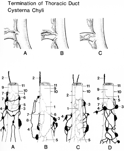
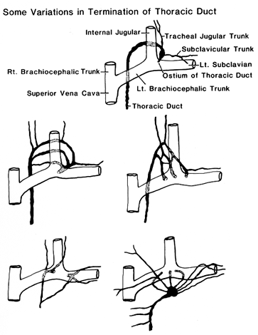
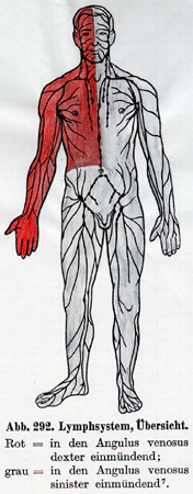
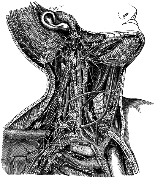
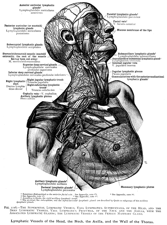
Lizars, John. A system of anatomical plates of the human body. Lymphatics of the head and neck. Published in Edinburgh about 1840. The right lymphatic duct forms various combinations with the right subclavian vein and jugular trunks. It is rare to find a simple right lymphatic duct that enters directly into the junction of the internal jugular and subclavian veins. The thoracic duct, which drains most of the body, may end by joining the left internal jugular, the junction of the left internal jugular and left subclavian vein, or the brachiocephalic vein.
Lizars, John. A system of anatomical plates of the human body. Lymphatics of the head and neck. Published in Edinburgh about 1840.
The right lymphatic duct forms various combinations with the right subclavian vein and jugular trunks. It is rare to find a simple right lymphatic duct that enters directly into the junction of the internal jugular and subclavian veins.
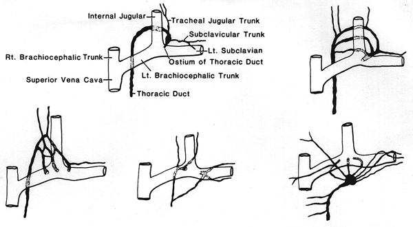
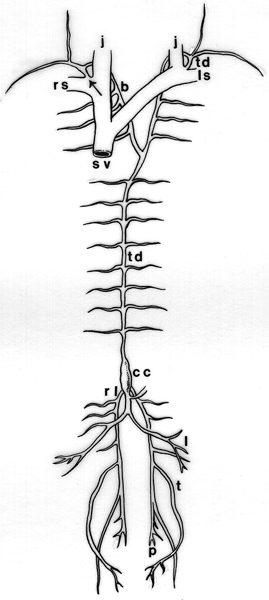
The thoracic duct, which drains most of the body, may end by joining the left internal jugular, the junction of the left internal jugular and left subclavian vein, or the brachiocephalic vein.
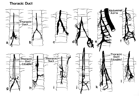
It is common for the duct to have several terminal divisions; it might be doubled or even tripled over a variable distance.
When doubled, it can open on both the right and left sides, at the venous angle.
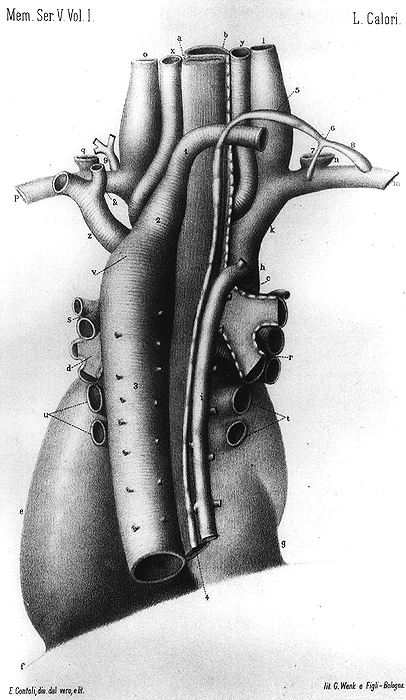
Persistence of paired ducts has been reported in cases of doubled aortic arch and in low origin (from the descending aorta) of the right subclavian.
Persistence of the (embryonic) right duct, with obliteration of the usual left duct, has been noted in a case of right aortic arch of the type presenting a left brachiocephalic trunk.
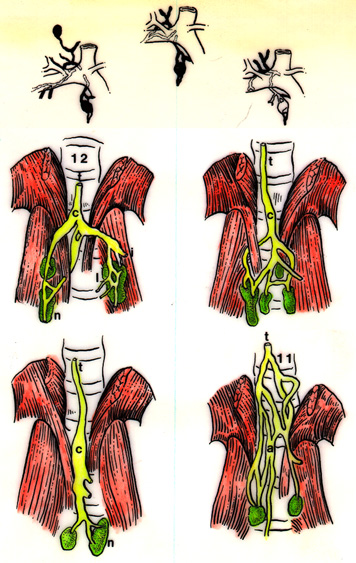
The size shape, and location of the cysterna chyli is extremely variable; the structure is absent in about 50% of cases examined.
The thoracic duct has been found in a normal subject, without other vascular variation, to terminate on the right side at the junction of the right subclavian and internal jugular veins.
The thoracic duct has also been found draining into the azygos vein and the inferior vena cava.
In one study, the thoracic duct opened into the following vessels (in order of decreasing frequency): a) left venous angle, b) internal jugular, c) left subclavian, d) vertebral, and e) others, including the azygos, hemiazygos and brachiocephalic. The thoracic duct has also been found draining into the subclavian artery.
A thoracic duct was found lying entirely to the left of the mid-vertebral line.
The length of the thoracic duct varies from 38 to 45 cm in the adult.
It usually begins at at the vertebral level of L2 but can begin at L3, L1, or even T12.
Anatomy Atlases. Immagine ospitata dal sito d’origine. Apri la pagina originale.
Anatomy Atlases. Immagine ospitata dal sito d’origine. Apri la pagina originale.
Anatomy Atlases. Immagine ospitata dal sito d’origine. Apri la pagina originale.
Anatomy Atlases. Immagine ospitata dal sito d’origine. Apri la pagina originale.
Anatomy Atlases. Immagine ospitata dal sito d’origine. Apri la pagina originale.
Anatomy Atlases. Immagine ospitata dal sito d’origine. Apri la pagina originale.
Anatomy Atlases. Immagine ospitata dal sito d’origine. Apri la pagina originale.
Anatomy Atlases. Immagine ospitata dal sito d’origine. Apri la pagina originale.
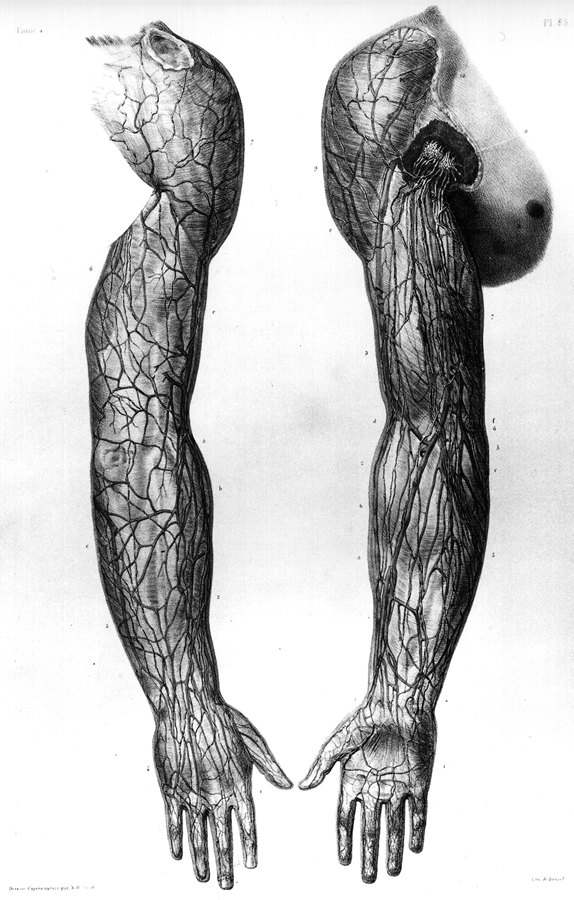
Anatomy Atlases. Immagine ospitata dal sito d’origine. Apri la pagina originale.
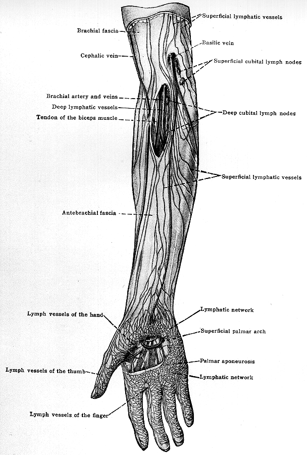
Anatomy Atlases. Immagine ospitata dal sito d’origine. Apri la pagina originale.
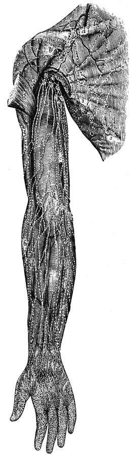
Anatomy Atlases. Immagine ospitata dal sito d’origine. Apri la pagina originale.
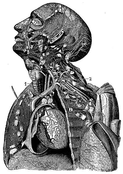
Anatomy Atlases. Immagine ospitata dal sito d’origine. Apri la pagina originale.
Anatomy Atlases. Immagine ospitata dal sito d’origine. Apri la pagina originale.
Anatomy Atlases. Immagine ospitata dal sito d’origine. Apri la pagina originale.
Riferimenti dell’opera originale
Elenco riportato come compare nella fonte, senza normalizzazione dei formati bibliografici.
- Bergman, R.A., Thompson, S.A., Afifi,A.K. F.A. Saadeh. (1988) Compendium of Human Anatomic Variation: Catalog, Atlas and World Literature. Urban & Schwarzenberg, Baltimore and Munich.
- Butler, C.S. (1903) On an abnormal thoracic duct. The Journal of Medical Research 10:153-156.
- Calori, L. (1880) Di una inversione splancnica generale nell'uomo accompagnata da alcuni notabili del capo con esso lei convenienti e da estranee anomalie. Mem. R. Accad. Sci. Istituto di Bologna. S. 4. 2:597-622.
- Calori, L. (1890) Sopa un caso d'inversione dei condotti toraicici accompagnato da inversa origine dell' arteria succlavia destra e sulla genesi della due anomalie. Mem. R. Accad. Sci. Istituto di Bologna. S.5 1:189-196.
- Davis, H.K. (1915) A statistical study of the thoracic duct in man. Am. J. Anat. 17:211-244.
- Elizarowski, S.I. (1941) Die chirurgische Anatomie des Halssbschnittes des Ductus thoracicus. Anat. Anz. 91:248-258.
- Golub, D.M. (1929) Ein Fall eines anomalen Ursprungs der A. subcl. dextra unterhalb der A. subcl. sin kombiniert mit dem Tr. bicaroticus und einem rechtsseitigen Münden des Ductus thoracicus. Anat. Anz. 67:387-392.
- Gortat-Zalewska, W. (1975) Variation of origin and course of the abdominal portion of the thoracic duct in man. Folia Morphol., Warsaw 34:53-58.
- Hafferi, A. (1923-24) Ein Fall von abnormen Verlauf des Ductus thoracicus und der hemiazygos. Anat. Anz. 57:373-378.
- Jakubczik, I. und H. Zeigler. (1963) Seltene Anomalien der grossen intrathoracalen Blut- und Lymphgefässstämme. Anat. Anz. 112:257-268.
- Jdanov, D.A. (1959) Anatomie du thoracique et des principaux collecteurs lymphatiques du tronc chez l'homme. Acta Anat. 37:20-47.
- Jossifow, G.M. (1905) Der Afang des Ductus thoracicus und desen Erweiterung. Arch. Anat. Physiol. Wissen Med. 1905:68-76.
- Llorca, O. (1935-36) über den rechtsseitigen Verlauf des Ductus thoracicus bei erhaltener rechter Aortenwurzel. Anat. Anz. 81:283-290.
- Minkin, A. (1925-26) Zur Frage des rechtsseitigen Verlaufes des Ductus thoracicus. Anat. Anz. 60:314-318.
- Nathan, H. and M.R. Seidel. (1983) The association of a retroesophageal right subclavian artery, a right-sided terminating thoracic duct, and a left vertebral artery of aortic origin: Anatomical and clinical considerations. Acta Anat. 117:362-373.
- Rodriguez, A.L. and J. M. Genis-Galvez. (1957) Varieté du ductus thoracicus non décrite chez l'homme: canal thoracique traversant l'anse Vieussens. Acta Anat. 31:61-65.
- Shepherd, F.J. (1882) On some anatomical variations: Common iliac arteries. Annals of Anatomy and Surgery. 6;172.
- Svitzer, -. (1845) Beobachtung einer Theilung des Ductus thoracicus. Arch. Anat. Physiol. Wissen. Med. 1845:21.
- Szawlowski, J. (1888) über das Verhalten des Ductus thoracicus bei Persistenz der rechten absteigenden Aortenwurzel. Anat. Anz. 3:839-849.
- Thomson, A. (1884) Variations of the thoracic duct associated with abnormal arterial distribution. J. Anat. Physiol. 18:416-425.
- Van Pernis, P.A. (1949) Variations of the thoracic duct. Surgery 26:806-809.
- Watson, M. (1872) Note on the termination of the thoracic ducton the right side. J. Anat. Physiol. 6:427-428.
- Wutzer, C.W. (1834) Einmünding des Ductus thoracicus in die Vena azygos. Arch. Anat. Physiol. 1834:311- 318.
- Walsham, W.J. (1880) The thoracic duct ending on the right side in the confluence of the internal jugular and subclavian veins. St Bartholomews Hospital Reports 16:99-100.
Classificazione anatomica
- Sistemi:
- Sistema cardiovascolare
Provenienza del contenuto
Adattamento
Adattamento di «Lymphatics: Thoracic Duct and Cysterna Chyli — Illustrated Encyclopedia of Human Anatomic Variation: Opus I: Muscular System» di Ronald A. Bergman, Adel K. Afifi, Ryosuke Miyauchi. Fonte: https://www.anatomyatlases.org/AnatomicVariants/Cardiovascular/Text/Lymphatics/ThoracicDuct.shtml Opera originale distribuita con licenza CC BY-NC-SA 4.0 (https://creativecommons.org/licenses/by-nc-sa/4.0/deed.it). Adattamento a cura di Redazione AIVA. Modifiche rispetto all’originale: Testo originale inglese riprodotto integralmente e riorganizzato nel formato scheda del portale (sintesi, testo, riferimenti), senza modificarne le affermazioni. La traduzione italiana non è ancora stata eseguita, e la pagina lo dichiara. Le figure non sono state copiate: sono collegate alla pagina d’origine. Questa pubblicazione non è approvata né avallata dagli autori dell’opera originale.
Licenza
Questo contenuto è distribuito con licenza CC BY-NC-SA 4.0. Licenze e attribuzioni