Voce enciclopedica
Arterie succlavie
Abstract
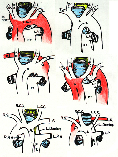
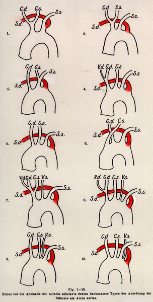
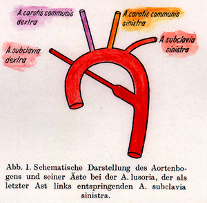
L’arteria succlavia destra può originare direttamente dall’arco aortico come primo, secondo, terzo, quarto o persino quinto ramo; quando nasce come ultimo ramo può decorrere dietro la trachea o dietro trachea ed esofago (arteria lusoria), comprimendo l’esofago e determinando la cosiddetta «disfagia lusoria». Sono descritte inoltre variazioni di rapporto con i muscoli scaleni, divisioni alte del vaso e, nei casi di arco aortico destro, un decorso retroesofageo della succlavia sinistra.
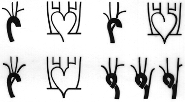
Le variazioni dell’arteria succlavia sono considerate anche insieme alle variazioni dell’arco aortico (elencate alla voce AORTA). Le irregolarità dei rami principali dell’arco aortico sono state classificate da Poynter come segue: 1). rami in numero inferiore al consueto, come nel caso della presenza di arterie bi-anonime; 2). rami in numero superiore al consueto, come nell’assenza dell’arteria anonima e nella conseguente origine separata di entrambe le arterie carotide e succlavia; 3). rami in successione anomala, dipendente da uno sviluppo irregolare del consueto piano dell’arco aortico. I casi qui riportati appartengono all’ultimo gruppo citato (3).
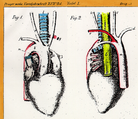
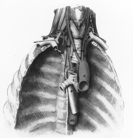
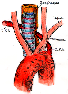
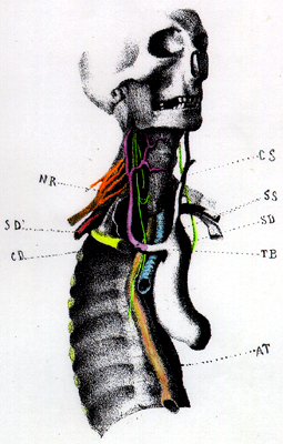
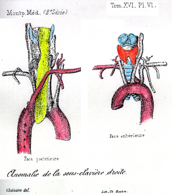
L’arteria succlavia destra può originare direttamente dall’arco dell’aorta, oppure come primo, secondo, terzo, quarto o persino quinto ramo di tale vaso. Quando origina come primo ramo, occupa il posto solitamente occupato dal tronco brachiocefalico. Quando origina come ramo distale o ultimo, può decorrere dietro la trachea (retrotracheale) o dietro sia la trachea sia l’esofago (retroesofagea) per raggiungere il solco sulla prima costa. Hommel (1737) descrisse il primo reperto autoptico di arteria succlavia destra originante come ultimo ramo dell’arco aortico. Nel 1789, ma senza che il caso fosse riportato prima del 1794, Bayford descrisse il primo paziente e la successiva autopsia di questo paziente con tale inusuale arteria succlavia destra. Cairney (1925) riassunse i risultati di otto ricercatori che avevano riportato 19 casi di questa variazione in 2291 soggetti autoptici (~0,8%). In quattro studi su 1783 corpi, la frequenza di questa variazione era di circa lo 0,6%. Il primo caso diagnosticato in vita mediante roentgenografia fu riportato da Kommerell nel 1936. L’incidenza riportata dagli studi radiologici è molto inferiore a quella degli studi autoptici (20/15000, ovvero ~0,13%). Haesemeyer e Gavant hanno riferito che nel loro studio radiologico su 7174 pazienti 36 presentavano un’arteria succlavia destra aberrante, con una frequenza dello 0,5%.
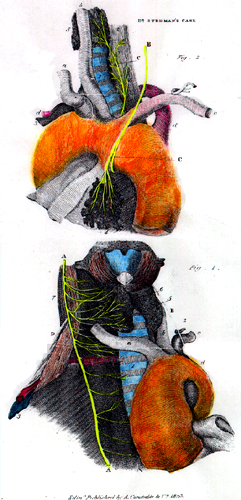
In altri casi, nei quali la succlavia destra è l’ultimo ramo dell’arco aortico, essa può passare fra la trachea e l’esofago oppure può seguire un decorso pretracheale per raggiungere il lato destro. Talvolta, quando l’arteria succlavia passa fra la trachea e l’esofago, ne deriva una compressione sia dell’arteria sia dell’esofago, che dà origine a una «malattia» denominata «disfagia lusoria» da Bayford (1794), dal termine «lusus naturae», che significa «scherzo di natura». Segue un estratto dalla memoria di Bayford: «Jane Fordham nacque a Bassenbourn, presso Royston, nell’Hertfordshire, nell’ultimo anno del secolo scorso (nota dell’autore: 1699). Fin dall’infanzia si osservò che aveva qualche difficoltà a deglutire, ma non vi si prestò molta attenzione finché non entrò nel tredicesimo anno di età, quando sperimentò per la prima volta quei sintomi che comunemente precedono la comparsa delle mestruazioni. In quel periodo la difficoltà divenne tanto considerevole da indurla a ricorrere alla medicina per averne sollievo; ma non trasse alcun beneficio apprezzabile da nulla, eccetto che da ripetuti salassi.... Proseguì in questo modo, con la malattia che aumentava gradualmente per molti anni, durante i quali osservò costantemente che la difficoltà di deglutire aumentava con l’esercizio violento e, come ella stessa si esprimeva, con tutto ciò che le riscaldava il sangue; sicché era spesso costretta a farsi salassare una o due volte negli intervalli del mese.... Negli ultimi vent’anni della sua vita questa povera creatura riusciva a stento, di giorno in giorno, a farsi forza per spingere giù il cibo ed evitare di morire di fame, tanto la difficoltà era ormai aumentata.
Descriveva questa difficoltà come derivante da un ostacolo posto in corrispondenza del primo osso dello sterno. Il cibo non refluiva quando giungeva in quel punto, ma sembrava subire un arresto momentaneo; e in quell’istante ella avvertiva un indescrivibile qualcosa di simile allo strangolamento o al soffocamento, che poteva paragonare soltanto a ciò che immaginava fossero le agonie della morte..... I diversi tipi di alimenti non producevano differenze apprezzabili negli effetti, salvo che i solidi le davano minore disagio dei liquidi: per questa ragione assumeva questi ultimi con grande parsimonia. Poiché i suoi disturbi venivano continuamente aggravati, anziché attenuati, dal tempo, ella divenne infine incapace di lottare; e, sfinita dalla fatica e dalla fame (si afferma infatti che negli ultimi tre settimane di vita a stento inghiottì un solo boccone), scese nella tomba all’inizio del febbraio 1761. Ritenni interessante indagare la causa dell’ostruzione mediante l’esame del cadavere. Non si giudicò necessario aprire il capo, poiché non vi era ragione di attendersi alcuna alterazione in quella parte. Furono esaminate a fondo le cavità del torace e dell’addome.... Alla fine, per puro caso, scoprii uno straordinario lusus naturae nella disposizione dell’arteria succlavia destra. La succlavia destra originava dalla parte posteriore dell’aorta, dietro e a sinistra della succlavia sinistra. Nel passare da sinistra a destra essa... si insinuava fra la trachea e l’esofago. Sembrerebbe che questa peculiare origine e decorso dell’arteria succlavia destra siano finora sfuggiti all’osservazione degli anatomici, altrimenti con ogni probabilità sarebbero stati registrati.... È impossibile supporre che l’atto della deglutizione si compia in questo caso senza un certo grado di pressione sull’arteria, là dove essa passa fra la trachea e l’esofago. Da ciò deve derivare un’interruzione della circolazione, la quale, in un’arteria così grande e così vicina al cuore, difficilmente potrebbe non suscitare sensazioni assai sgradevoli.... L’atto della deglutizione non sarà accompagnato da dolore o indolenzimento, bensì da estrema angoscia e da violente palpitazioni del cuore». Qui termina la relazione di Bayford.
La succlavia è raramente il secondo o il terzo ramo dell’arco aortico, ma quando lo è decorre dietro la carotide comune destra.
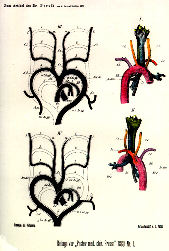
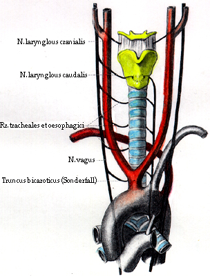
La succlavia destra origina, dal punto di vista embriologico, come ramo distale o ultimo dell’arco aortico quando l’arco aortico destro rimane integro e la normale radice dell’arteria succlavia si è obliterata. Un’arteria aberrans, originante dalla succlavia destra (o dall’intercostale superiore), può in genere essere seguita fino alla terza vertebra toracica dietro l’esofago e, in un certo numero di tali casi, può essere seguita attraverso la colonna vertebrale, dove si anastomizza con un ramo dell’aorta toracica che origina al di sotto del dotto arterioso. È l’ampliamento di questa anastomosi (che rappresenta il residuo dell’aorta dorsale destra primitiva dell’embrione) a dare origine alla variazione. In tali casi il nervo laringeo ricorrente segue un decorso diretto verso la laringe, invece di girare in modo ricorrente attorno all’arteria succlavia, perché il quarto arco destro, che forma la prima porzione della succlavia, è obliterato.
La succlavia destra può originare più in alto o più in basso nel collo rispetto al consueto, a seconda che il tronco brachiocefalico si divida al di sopra o al di sotto della sua posizione normale. In alcuni casi la succlavia origina dall’aorta toracica.
La succlavia può perforare il muscolo scaleno anteriore o passare davanti ad esso; quando perfora lo scaleno anteriore si possono formare anelli arteriosi attorno a linguette muscolari. L’arteria può risalire al di sopra del livello della clavicola o rimanere al di sotto di esso. La terza porzione dell’arteria può essere coperta dal muscolo trapezio o dallo sternocleidomastoideo, oppure dall’origine clavicolare del muscolo omoioideo. La vena succlavia accompagna occasionalmente l’arteria dietro lo scaleno anteriore; l’arteria può anche passare fra lo scaleno medio e il posteriore. L’arteria succlavia destra può ricevere il dotto toracico; la succlavia sinistra può ricevere un dotto arterioso pervio.
In alcuni casi l’arteria succlavia si divide in corrispondenza del margine mediale del muscolo scaleno anteriore, e i due rami proseguono attraverso l’ascella e lungo il braccio per diventare le arterie radiale e ulnare.
Le arterie succlavie destre anomale ricorrono nello 0,4-1,7% dei soggetti (secondo tutte le fonti). Quain ha riportato 4 casi/1000, Holzapfel 6/1000 e Tiedemann 8 volte su 1000 casi. Goldbloom ha riportato 4/225 cadaveri (1,7%), Harvey (U. Cal. Medical School) ne ha trovati 2/237 (0,8%), Cobey ha riportato in Thomson’s Collective Investigation of the Anatomical Society of Great Britain and Ireland 5/500 (1%). È interessante che il decorso dell’arteria succlavia fosse retroesofageo (fra la vertebra e l’esofago) nella maggior parte dei casi (107/238), retrotracheale (fra la trachea e l’esofago) di rado (25/238 casi) e davanti alla trachea molto raramente (6/238 casi).
Se è presente una costa cervicale, l’arteria può decorrere al di sopra di questa costa soprannumeraria e inusuale.
Nei casi di arco aortico destro, l’arteria succlavia sinistra può essere retroesofagea quando origina come ultimo ramo dell’arco. La succlavia sinistra può essere doppia e originare come primo e come quinto ramo dell’arco aortico. L’ultimo ramo è retroesofageo.
Nei casi di arco aortico destro, la succlavia sinistra mostra due variazioni principali: 1) quando la succlavia sinistra origina come primo grosso vaso dell’arco aortico destro, essa incrocia la faccia anteriore della trachea per raggiungere il lato sinistro; 2) quando l’arteria succlavia sinistra origina come ultimo ramo dell’arco aortico, essa incrocia la faccia posteriore dell’esofago per raggiungere il lato sinistro. La succlavia sinistra non origina direttamente dall’arco aortico, bensì da un diverticolo, residuo del quarto arco branchiale sinistro.
Holzapfel, che studiò il decorso di 133 casi di succlavia destra originante come ultimo ramo dell’arco aortico, trovò che in 107 casi (80%) la succlavia destra passava dietro l’esofago per raggiungere il lato destro, fra la trachea e l’esofago in 20 casi (15%) e davanti alla trachea in 6 casi (5%). Banchi ha riferito su 165 casi: 138 casi (83,6%) dietro l’esofago, 21 casi (12,7%) fra trachea ed esofago e 7 casi (4,2%) davanti alla trachea. Cairney, nel 1925, riassunse i dati di otto autori e riportò 19 casi di arteria succlavia destra variabile in 2291 reperti autoptici. Altri hanno riportato un’incidenza dallo 0,4% al 2,0% negli studi anatomici. Il primo caso diagnosticato in un paziente vivente mediante roentgenologia fu riportato da Kommerell nel 1936, ma l’incidenza dei casi individuati con i raggi X è inferiore a quella delle casistiche autoptiche. Brombart trovò soltanto 20 casi (0,13%) su circa 15.000 soggetti.
Elenco alfabetico degli autori e delle date dei casi riportati di decorso inusuale dell’arteria succlavia destra quando questa origina come ultimo ramo dell’arco aortico – prima del 1900. Da Holzapfel, 1899.
Arnold, 1847.
Brodie, 1888.
Autenrieth-Pfleiderer, 1806, 1807.
Brown, 1868.
Bankart, 1869.
Cassebohm-Böhmer, 1741.
Barkow, 1866, 1869.
Cavasse, 1856.
Barwell, 1867.
Cerutti, 1827.
Bayford, 1789.
Colles, 1811.
Blandin, 1848.
Cruveilhier, 1832.
Böhmer, 1741.
Demarquay, 1848.
Bothezat, 1891.
Demeaux, 1841.
Bradley, 1781.
v. Düben, 1876.
Brenner, 1883.
Dubrueil, 1837, 1847, 1862.
Brent, 1844.
Dubrueil-Sappey, 1847.
Brewer, 1791.
Dunn, 1890.
Erdmann, 1772.
Löseke, 1754.
Faure, 1895.
Ludwig, 1764.
Fleischmann, 1815, 1835.
Macartney-Tiedemann, 1846.
Flesch, 1879.
Mayer, 1827.
Frandsen, 1854.
Mears, 1871.
Giacomini, 1882.
Meckel, 1751, 1805, 1810, 1816, 1820.
Godman, 1824.
Mieg, 1753.
Goetz, 1896.
Monro, 1797.
Green, 1830.
Murray, 1771.
Gross-Mears, 1871.
Neubauer-Erdmann, 1772.
Harrison, 1839.
Oehl, 1859.
Hart, 1826.
Otto, 1816,1830.
Hart-Quain, 1844.
Patruban, 1844.
Herold, 1812.
Peacock, 1860.
Hesselbach, 1824.
Pfleiderer, 1806, 1807.
Hoffman-Fabricius, 1751.
Pigné, 1847.
Hommel, 1737.
Pohl, 1773.
Hopkinson, 1830.
Pye-Smith, 1871.
Hulme, 1789.
Quain, 1844.
Hunauld, 1735. (Primo caso riportato)
Rau, 1890.
Hyrtl, 1841-1859.
Reid, 1846.
Isenflamm-Fleischmann, 1800, 1845.
Sandifort, 1772, 1793.
Jacques, 1895.
Sappey-Dubrueil, 1847.
Kirby, 1818.
Schleitz, 1771.
Koberwein, 1810.
Shepherd, 1889.
Krause, 1876.
Simon, 1846.
Lauth, 1830.
Smith, 1891. (Kaninchen = coniglio)
Leboucq, 1894.
Solger, 1893.
Leidy-Mears, 1871.
Stachelroth, 1850.
Lenoir, 1832.
Stedman, 1823.
Liston, 1839.
Struthers, 1888.
Testut, 1896.
Walsham, 1880.
Thomson, 1890.
Walter, 1785.
Tiedemann, 1822, 1846.
Weber, 1829.
Turner, 1862.
Wood, 1859, 1867.
Valentin, 1791.
Zagorsky, 1810.
Wagner, 1828.
Zenker, 1878.
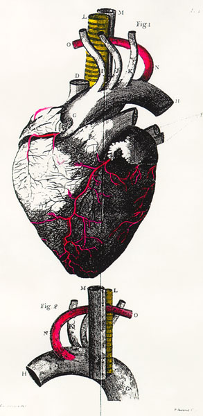
Retroesofagea sinistra
Anatomy Atlases. Immagine ospitata dal sito d’origine. Apri la pagina originale.
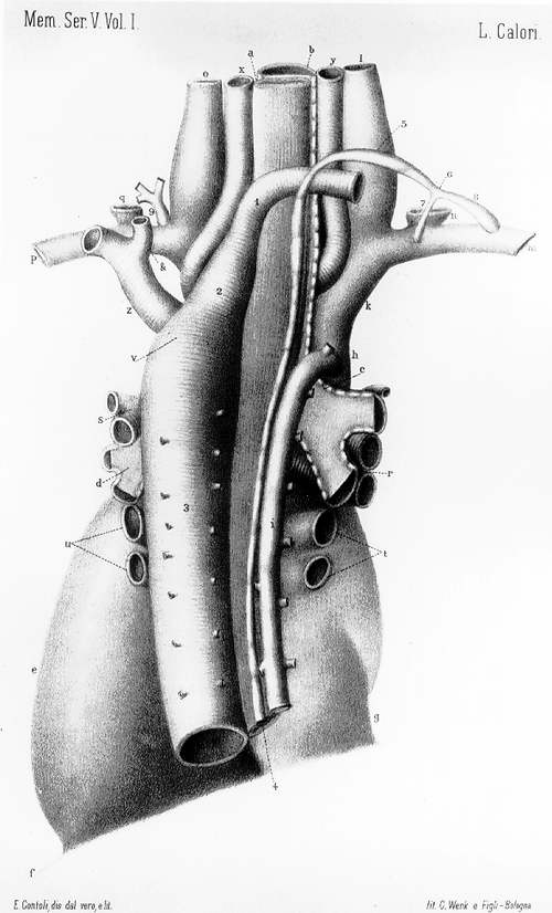
Retroesofagea sinistra
Anatomy Atlases. Immagine ospitata dal sito d’origine. Apri la pagina originale.
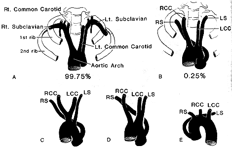
Retroesofagea sinistra
Anatomy Atlases. Immagine ospitata dal sito d’origine. Apri la pagina originale.
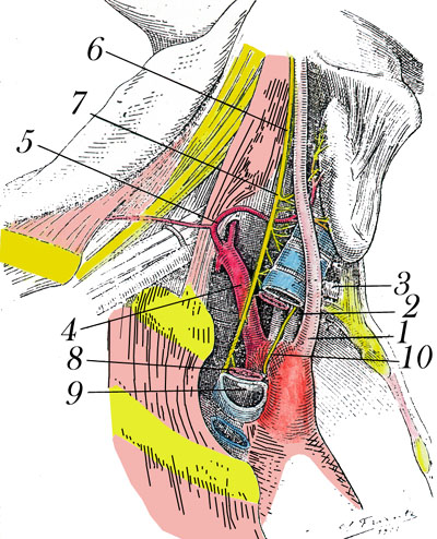
Retroesofagea sinistra
Anatomy Atlases. Immagine ospitata dal sito d’origine. Apri la pagina originale.
Destra
Anatomy Atlases. Immagine ospitata dal sito d’origine. Apri la pagina originale.
Destra
Anatomy Atlases. Immagine ospitata dal sito d’origine. Apri la pagina originale.
Destra
Anatomy Atlases. Immagine ospitata dal sito d’origine. Apri la pagina originale.
Vedi , , , , , , , , , , , , , , Vedi , , ,
Anatomy Atlases. Immagine ospitata dal sito d’origine. Apri la pagina originale.
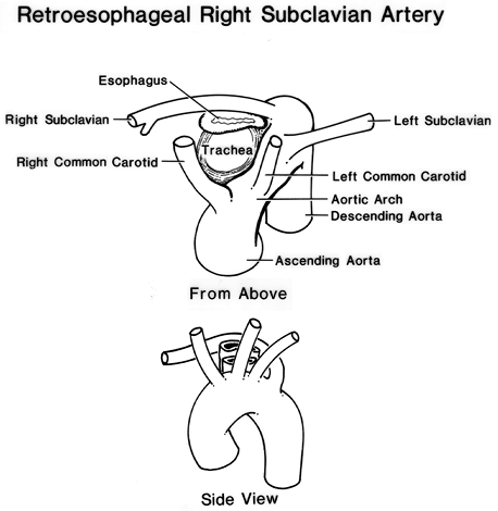
Anatomy Atlases. Immagine ospitata dal sito d’origine. Apri la pagina originale.
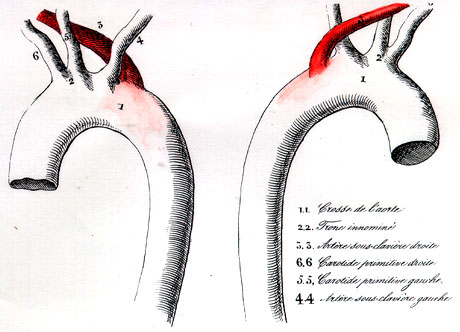
Anatomy Atlases. Immagine ospitata dal sito d’origine. Apri la pagina originale.
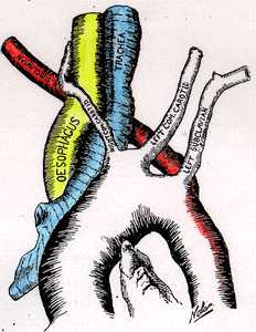
Anatomy Atlases. Immagine ospitata dal sito d’origine. Apri la pagina originale.
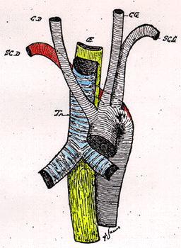
Anatomy Atlases. Immagine ospitata dal sito d’origine. Apri la pagina originale.
Anatomy Atlases. Immagine ospitata dal sito d’origine. Apri la pagina originale.
Anatomy Atlases. Immagine ospitata dal sito d’origine. Apri la pagina originale.
Anatomy Atlases. Immagine ospitata dal sito d’origine. Apri la pagina originale.
Anatomy Atlases. Immagine ospitata dal sito d’origine. Apri la pagina originale.
Anatomy Atlases. Immagine ospitata dal sito d’origine. Apri la pagina originale.
Anatomy Atlases. Immagine ospitata dal sito d’origine. Apri la pagina originale.
Anatomy Atlases. Immagine ospitata dal sito d’origine. Apri la pagina originale.
Anatomy Atlases. Immagine ospitata dal sito d’origine. Apri la pagina originale.
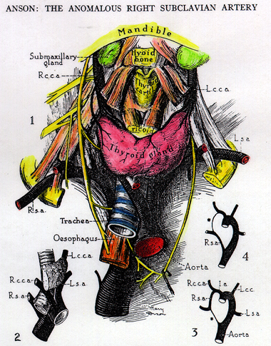
Anatomy Atlases. Immagine ospitata dal sito d’origine. Apri la pagina originale.
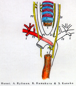
Anatomy Atlases. Immagine ospitata dal sito d’origine. Apri la pagina originale.
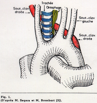
Anatomy Atlases. Immagine ospitata dal sito d’origine. Apri la pagina originale.
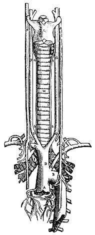
Anatomy Atlases. Immagine ospitata dal sito d’origine. Apri la pagina originale.
Retrotracheale
Vedi immagine 43
Arteria lusoria dextra (destra) e arteria subclavia dextra lusoria
Vedi immagini 230 , 231 , 394
Arteria lusoria sinistra (sinistra)
Vedi immagine 425
Riferimenti dell’opera originale
Elenco riportato come compare nella fonte, senza normalizzazione dei formati bibliografici. Le voci non sono tradotte: un riferimento bibliografico si cita nella lingua in cui è pubblicato.
- Abbott, M. (1936) Atlas of Congenital Cardiac Disease. American Heart Assoc., New York.
- Anile, A. (1898) Di un'importante anomalia artero - nervosa (arteria succlavia di destra e nervo laryngeo inferiore corrispondente). La Riforma Medica 2:411-416.
- Anon., (1791) Origine bizarre de I'artère sous-clavière. J. de Medecine, Chirurgie, Pharmacie, Dc. 86:239-241.
- Anson, B.J. (1936) The anomalous right subclavian artery. Surg., Gynecol. Obstet. 62:708-711.
- Arendt, J. and A. Wolf. (1947) The vallecular sign: Its diagnosis and clinical significance. Am. J. Roentgenology (AJR) 57:435-445.
- Autenrieth, -. (1807-08) Zusatz zu der Abhandlung: De Dysphagia lusoria. Arch. Physiol. 8:264-268.
- Autenrieth, -. und -. Pfleiderer. (1807) Disseratatio inauguralis. De Dysphagia lusoria. Arch. Physiol. 7:145-188.
- Bayford, D. (1794) An account of a singular case of obstructed deglutition. Mem. Med. Soc. Lond. 2:271-286.
- Beabout, J.W., Stewart, J.R. and O.W. Kincaid. (1964) Aberrant right subclavian artery: dispute of commonly accepted concepts. Am. J. Roentgenol. (AJR) 92:855-864.
- Bean, R.B. (1904) Observations on a study of the subclavian artery in man. Johns Hopkins Hosp. Bull. 15:203-205.
- Bean, R.B. (1905) A composite study of the subclavian artery in man. Am. J. Anatomy 4:303-328.
- Beaumanoir (de Brest), -. (1882) Présentation de pièces anatomiques: Sous-clavière. Bull. de la Soc. Anatomique de Paris 57:318-320.
- Beaumanoir, (de Brest.), -. (1882) Anomalies artèrielles trouvées a l'autopsie d'un sujet. Bull. de la Soc. Anatomique de Paris 57:316-321.
- Bereza, S. (1966) A case of aberrant subclavian artery and other neurovascular anomalies in the neck. Folia Morphol., Warsaw 25:589-599.
- Bergman, R.A., Thompson, S.A., Afifi, A.K. and F.A. Saadeh. (1988) Compendium of Human Anatomic Variation: Catalog, Atlas and World Literature. Urban & Schwarzenberg., Baltimore and Munich.
- Bevier, G. (1915) An anomalous origin of the right subclavian artery. Anat. Rec. 9:785-789.
- Billet, H. (1937) Suite de l'étude des rapports des branches collatérales de la sousclavière avec les troncs primaires du plexus brachial. Assoc. Anatomistes Comptes Rendus 32:49-52.
- Blair, D.M., Davies, F. and W. McKissock. (1934-35) The etiology of the vascular symptoms of cervical rib. Br. J. Surg. 22:406-414.
- Blalock, A. (1947) Technique of creation of an artificial ductus arteriosus in the treatment of pulmonary stenosis. J. Thoracic Surgery 16:244-254.
- Blumgart, H.L. and A.S. Freedberg. (1955) Arterial malformations which cause compression of the trachea or esophagus. Circulation 11:133-134.
- Bochdalek, Jr., -. (1867) Einsenkung des Lig. arteriosum (Ductus arteriosus Botalli) in die linke Arteria subclavia. Arch. Pathol. Anat. Physiol. Klin. Med. 41:259-260.
- Bothezat, P. (1891) Anomalie de l'artère sous-clavière droite. Montpellier Médical Series 2, 16:418-420.
- Bouchet, P. (1903) Anomalie de naissance et de trajet de l'artère sous-clavière droite. Bull. et Mém. de la Soc. Anatomique de Paris. 5:155.
- Boyd, G.I. (1934) Abnormality of subclavian artery associated with presence of the scalenus minimus. J. Anat. 68:280-281.
- Bradley, S.M. (1871) Note on a peculiar origin of the right subclavian artery. J. Anat. Physiol. 5:341-342.
- Branscom, J.J. and J.H.M. Austin. (1973) Aberrant right subclavian artery. Findings seen on plain chest roentgenograms. American Journal of Roentgenology, Radium Therapy and Nuclear Medicine (AJR) 119:539-542.
- Brean, H.P. and E.B.D. Neuhauser. (1947) Syndrome of aberrant right subclavian artery with patent ductus arteriosus. Am. J. Roentgenology (AJR) 58:708-716.
- Brewer, -. (1791) Observation, par M. Bayford. Déglutition empêchée par la position extraordinaire de l'artère sous-claviere. J. de Chirurgie 2:281.
- Brombart, M. et J. Schuemans (1951) Pathologie meconnue de l'esophage. Acta O.R.L. Belgica 5:5-45.
- Buschendorff, C. (1917) Beitrag zur Kenntnis der Persistenz eines doppelten Aortenbogens und seine klinische Bedeutung. Zentralblatt für Herz- und Gefasskrankheiten 9:165-171.
- Cairney, J. (1924-1925) The anomalous right subclavian artery considered in the light of recent findings in arterial development; with a note on two cases of an unusual relation of the innominate artery to the trachea. J. Anat. 59:265-296.
- Calori, L. (1868) Delle anomalie piu importanti di ossa, vasi, nervi, e muscoli occorse nell'ultimo biennio facendo anatomia del corpo umano. Mem. della Accademia delle Scienze dell'Istituto di Bologna, S.2. 8:417-482.
- Calori, L. (1890) Sopra un caso d'inversione dei condotti toracici accompagnato da inversa origine dell'arteria succlavia destra e sulla genesi della due anomalie. Mem. R. Accad. Sci. Istituto di Bologna, S.5. 1:189-196.
- Carrier, A.E. (1880) Pecularities in the origin and course of Arteries of the upper extremities. Toledo Med. Surg. J., Ohio 4:365-367.
- Casali, R. (1911) Di tre rami soprannumerari non ancora descritti dell'A. succlavia. Arteria del muscolo succlavio, Arteria della cupola pleurica, Arteria esfogea superiore. Arch. Ital. Anat. Embriol. 10:122-128.
- Casali, R. (1912) Osservazioni sui rami collaterali dell'a succlavia. Bibliographie Anatomique 22(2):41.
- Chacón, F. de P. (1892) Anomalia de la arteria subclavia. Gaceta Médica de Mexico 28:442-446.
- Cobey, J.F. (1914) An anomalous right subclavian artery. Anat. Rec. 8:15-19.
- Coplemann, B. (1945) Anomalous right subclavian artery. Am. J. Roentgenol. (AJR) 54:270-275.
- Costa, A.J. (1946) A proposito de una anomalia arterial. La arteria subclavia derecha saliendo del cayado aortico en su porción final. Rev. Assoc. Méd. Argentina 60:740-743. Cited in Excerpta Medica, Sec. 1, Vol. 2, abstract 525, p.250, 1948.
- Coues, E. (1880) Anatomical anomalies. The Medical Record, N.Y. 17:257.
- Craig, W.B. (1880) Unusual origin of the subclavian artery. Gaillards Medical Journal 30:428-429.
- Curnow, J. (1888) Right subclavian artery passing in front of the scalenus anticus. Proc. Anat. Soc. of Great Britain and Ireland. J. Anat. Physiol. 22:v-vi.
- Dahm, M. (1940) Zür Eindellung der Spieseröhre bei links entspringender Arteria subclavia dextra. Fortschr. Röntgenstr. 62:108-114.
- Dalton, A. and W.F. Alexander. (1947) Anomalous right subclavian artery originating from the descending aorta. Anatomical Record 97:328.
- Daseler, E.H. and B.J. Anson. (1959) Surgical anatomy of the subclavian artery and its branches. Surg., Gynecol. Obstet. 108:149-174.
- Davies-Colley, N., Taylor, F. and B.N. Dalton. (1873) Left carotid arising from the innominate. Guy's Hosp. Reports 18:389-401.
- Deaver, J.B. (1888/89) Anomalies of the arch of the aorta. Univ. Med. Mag., Philadelphia 2:40-41.
- DeGaris, C.F. (1924) Patterns of branching of the subclavian artery in white and negro stocks. Am. J. Phys. Anthrop. 7:95-107.
- DeGaris, C.F. (1932) Aortic axillary collaterals and the pattern of arm arteries in anomalous right subclavian artery. Am. J. Anat. 51:189-213.
- Deniker, M. (1913) Anomalies d'origine et de trajet des branches de la crosse aortique. Bull. et Mem. de la Soc. Anatomique de Paris. 88:127-129.
- Denisoff, B.N. (1912) Abnormal origin of the right subclavian artery. (In Russian) Russkii Vrach 11:1069-1071.
- Dolgopol, V.B. (1934) Anomalous origin of the right subclavian from the descending arch of the aorta. J. Tech. Methods 13:112-118.
- Dolgo-Saburoff, B. (1933) Zur Frage über die Lagebeziehungen zwischen der Arteria subclavia und Scalenusmuskulatur beim Menschen. Anat. Anz. 76:97:113.
- von Düben, G. (1876) Abnorm verlaufende Arteria subclavia dextra. Svenska Läkaresällskapet, Förhandlingar vid 38:81-82.
- Dubreuil, -. (1837) Déplacement et anomalies de rapport des veine et artère sous-clavières droites ayant mis obstacle a la ligature de ce dernier vaisseau.- Indication de quelques variétés dans l'origine et le trajet des artères sous-clavières. Gaz. Médicale de Paris 2th series, 5:562-565.
- Dubreuil, -. (1862) D'anomalie d'origine de l'artère sous-clavière droite. Bull. et Mém. de la Soc. Anatomique de Paris. 37:443.
- Dubreuil, -. (1862) Exemple d'anomalie d'origin de l'artère sous-clavière droit. Bulletins et Mem. de la Société Anatomique de Paris XXXVII(12):443.
- Dunn, L.A., Washburn, J.W. and J.H. Targett. (1890) Abnormalities observed in the dissecting room of Guy's Hospital during sessions 1888-1890. S. 3., 32:299-315.
- Eklöf, O., Ekström, G., Erikson, B.O. et al. (1971) Arterial anomalies causing compression of the trachea and/or the esophagus. Acta Pediatr., Scand. 60:81-89.
- Farlow, J. W. (1886) Abnormal artery of the pharynx. Boston Medical and Surgical Journal. CXIV(19):437-438.
- Faure, J. -L. (1895) Anomalie d'origine de l'artère sous-clavière droite. Bull. et Mem. de la Societe Anatomique de Paris 9:11-12.
- Felson, B., Cohen, S., Courter, S.R. and J. McGuire. (1950) Anomalous right subclavian artery. Radiology 54:349-349.
- Fischer, J. (1858) Verlauf der art, subclavia in einem Falle von deutlich entwickelten Halsrippen. Wiener Medizinische Wochenschrift 8 (No. 30) 24 Juli:537-539
- Fossei, H.M. (1942) Ueber Dysphagia lusoria. Fortschr Röntgenstr. 79(3):111-112.
- Freed, K. and V.H.S. and Low (1996) The aberrrant subclavian artery. AJR 168:481-484.
- Furukawa, H. and T. Takemurs. (1995) Stanford type A acute aortic dissection involving an aberrant right subclavian artery: a successful operative case. (In Japanese). Nippon Kyobu Geka Gakkai Zasshi 43:533-537.
- García Cañizares, D.F. Anomalias Arteriales. Revista de Ciencias Medicas 10: 75-77.
- Geddes, A.C. (1911) A note on abnormal right subclavian artery. J. Anat. Physiol. 45:197-198.
- Geddes, A.C. (1911) Note on the reputed passage of abnormal right subclavian artery (A) in front of the trachea, (B) between the trachea and oesophagus. J. Anat. Physiol. 45:199-201.
- Giannelli, L. (1894) Sopra alcune Anomalie dei vasi. Atti d. R. Accad. d. Fisiocritici in Siena. S.4. 6:363-370.
- Gladstone, R.J. and C.P.G. Wakeley. (1915) Two cases, considered from the developmental standpoint, in which the right subclavian artery arose from the arch of the aorta beyond the origin of the left subclavian artery; with a note on the relation of the subclavian veins to the cardinal system. J. Anat. Physiol. 49:362-374.
- Goldbloom, A.A. (1922) The anomalous right subclavian artery and its possible clinical significance. Surg., Gynecol. Obstet. 34:378-384.
- Golub, D.M. (1929) Ein Fall eines anomalen Ursprungs der A. subcl. dextra unterhalb der A. subcl. sin. kombiniert mit dem Tr. bicaroticus und einem rechtsseitigen Münden des Ductus thoracicus. Anat. Anz. 67:387-392.
- Gould, E.L.P. (1909) A case of abnormal right subclavian artery. J. Anat. Physiol. 43:324-328.
- Gross, C., Prebl, F. and P. Brucke. (1990) Thoracic aneurysm in association with an aberrant right subclavian artery. Eur. J. Cardiothoracic Surg. 4:105-106.
- Gross, R.E. (1946) Surgical treatment for dysphagia lusoria. Ann. Surg. 124:532-534.
- Gross, R.E. (1955) Arterial malformations which cause compression of the trachea or esophagus. Circulation 11:124-134.
- Gross, R.E. and E.B.D. Neuhauser. (1951) Compression of the trachea or esophagus by vascular anomalies. Surgical therapy in 40 cases. Pediatrics 7:69-88.
- Gross, R.E. and P.F. Ware. (1946) Surgical significance of aortic arch anomalies. Surg., Gynecol. and Obstet. 83:435-448.
- Guiria, P.M. (1890) Ancora del rapporto che si stabilisce fra l'A. vertebrale destro e il N. laringeô inferiore, quando ecc. Mem. della R. Accademia Medica di Genova 1890:7-17.
- Günsel, E. (1940) Dysphagia lusoria bei Arcus aortae dexter und sinister. Röntgenpraxis 12:346-348.
- Haesemeyer, S.W. and M.L. Gavant. (1999) Imaging of acute traumatic aortic tear in patients with aberrant right subclavian artery. AJR 172:117-120.
- Hamann, C.A. (1900) Right subclavian artery passing behind the trachea and esophagus - other anomalies. Cleveland J. Med. 5:453-455.
- Hammer, D.L. and A.M. Meis. (1941) Thyroid arteries and anomalous subclavian in the white and negro. Am. J. Phys. Anthropol. 28:227-237.
- Hanai, H., Ryúmon, A., Kamakura, K. and S. Kaneko. (1963-64) An anomalous case of A. Subclavia dextra rising from arcus aortae as the last branch. Okajimas Folia Anatomica Japonica 39:31-36.
- Hart, J. (1826) A case of irregular origin and course of right subclavian artery. Edinburgh Med. J. 25:286-287.
- Harvey, R.W. (1917) Notes on two cases of anomalous right subclavian artery. Anat. Rec. 12:329-330.
- Haug, H. and U. Merkel. (1965) Abnormer Ursprung und Verlauf der A. subclavia dextra. Anat. Anz. 117:319-321.
- Herbut, P.A. (1943) Anomalies of the aortic arch. Arch. Pathol. 35:717-729.
- Herringham, W.P. (1891) Right aorta with persistent left aortic root giving origin to the left subclavian. Proc. Anat. Soc. J. Anat. Physiol. 25:vi-vii.
- Holtzapfel, G. (1899) Ungewöhnlicher Ursprung und Verlauf der Arteria subclavia dextra. Anat. Hefte 12:369-523.
- Hopkinson, J.P. (1831) Aneurism of the aorta, and usual origin of right subclavian. Am. J. Med. Sci. 7:556-557.
- Hudson, W.A. (1921) A case of an anomalous right subclavian artery. Washington University Studies. IX, Scientific series 1:219-228.
- Hunauld, L. (1735) Examen de quelques parties d'un singe. Hist. Acad. Roy. Sci. 2:516.
- Jacques, P. (1895) Quelques anomalies artèrielles (sous-clavière droite, tronc coeliaque, poplitée). Bull. et Mém. de la Société Anatomique de Paris 70:252-255.
- Kalbfleisch, H.H. (1948) Durch morphologische Veränderungen nachweisbare funktionale Segmente in Pharynx, Hypopharynx und Oesophagus. Allgemeinpathol. Schriften: Beitr. zur allgemein. path. aus der Spezialen Path. 7:5-25.
- Kaplan, E.B. (1947) Variations of the subclavian vessels in the region of the scalenus anterior muscle. N.Y. Hosp. Joint Dis. Bull. 8:217-218.
- Kirby, J. (1818) A case in which suffocation was produced by a portion of solid food in the oesophagus. The Dublin Hospital Reports and Communications in Medicine and Surgery 2:224-226.
- Klinkhamer, A.C. (1966) Aberrant right subclavian artery. Clinical and roentgenologic aspects. Am. J. Roentgenology (AJR) 97:438-446.
- Kodama, K. (1935) Two cases of the right subclavian artery as the last branch of the aortic arch. Mitt. Med. Akad. Kioto 14:961-964.
- Kommerell, B. (1936) Verlagerung des ösophagus durch eine abnorm verlaufende Arteria subclavia dextra (Arteria lusoria). Fortschr. Röntgenstr. 54:590-595.
- Kroeker, E.J., Kirklin, J.W., Prickman, L.E. and E.H. Wood. (1954) Coarctation of the aorta, subaortic stenosis, anomalous right subclavian artery: Report of case. Staff Meetings of the Mayo Clinic 29(10):266-277.
- Krokowski, E. (1960) Arteria lusoria. Fortschr. Röntgenstr. 93:376-377
- Langman, J. (1954) Oesophageal atresia accompanied by a remarkable vessel anomaly. Archivum Chirurgicum Neerlandicum 4:39-42.
- Lombardi, G. (1936) Il quadro radiologico della arteria lusoria (arteria succlavia destra anomala). Radiol. Med. 37:809-816.
- Mears, J.E. (1871) Description of an anomalous origin of the right subclavian artery, associated with anomalies of origin of the branches of both subclavian arteries; with remarks. Am. J. Med. Sci. 62:401-409.
- Menke, J.F. (1936) Anomalous a. bronchialis dextra from the a. subclavia dextra, secondarily connected to the aorta thoracalis. Anat. Rec. 65:55-58.
- Michaud, -et A. Antoine. (1834) Anomalies dans la mode dorigine des branches de la crosse aortique. J. des Sciences Medicales de Montpellier 1:177-180.
- Milianitch, M.N. (1924) Note sure une anomalie d'origine des grostrones nés de la crosse de l'aorte accompagné d'une anomalie de trajet du nerf récurrent droit. bull. et Mém de la Soc. Anatomique de Paris. 94:230-233.
- Moreira da Rocha, J. (1926) Arteria subclavia direita como ultimo ramo da crossa aortica. Ann. da Faculd. de Med. de São Paulo, Brasil 1:103-124.
- Mouton, C. (1919) Ueber Anomalien der Arteria subclavia dextra und ihre Folgezustände (Dysphagia lusoria). Brun's Beiträge zur Klinischen Chirurgie 115:365-407.
- Nakagawa, M. (1938) On the right subclavian artery as the last branch of the aortic arch. (In Japanese) Juzenkai Z. 44(1):203-216.
- Nathan, H. and M.R. Seidel. (1983) The association of a retroesophageal right subclavian artery, a right-sided terminating thoracic duct, and a left vertebral artery of aortic origin: Anatomical and clinical considerations. Acta Anat. 117:362-373.
- Natsis, K., Didagelos, M., Gkiouliava, A. et al. Surg Radiol Anat (2016). doi:10.1007/s00276-016-1796-5
- Neuhauser, E.B.D. (1946) Roentgen diagnosis of double aortic arch and other anomalies of the great vessels. Am. J. Roentgenol. (AJR) 56:1-12.
- Nizankowski, C. Noczynski, L. and E. Suder. (1982) Variability of the origin of ramifications of the subclavian artery in humans (studies on the Polish population). Folia Morphol., Warsaw 41:281-294.
- O'Rahilly, R., Debson, H. and T.S. King. (1950) Subclavian origin of bronchial arteries. Anat. Rec. 108:227-238.
- Ozenne, -. (1883) 12. Anomalie d'origine de la carotide droite et de l'artère sous-clavière du même côté. Bull. de la Société Anatomique de Paris, 4th series, 8:108.
- Ozenne, -. (1883) 12. Anomalie d'origine de la carotide droite et de l'artère sous-clavière du même côté. Le Progrès Médical 11:734.
- Palmer, E.D. (1955) Dysphagia lusoria: Clinical aspects in the adult. Annals of Internal Medicine 42 (6):1173-1180.
- Papadatos, D. (1976) Trois observations anatomiques sur l'origine de l'arteria subclavia dextra comme denière branche de l'arcus aortae (arteria lusoria). Anat. Anz. 140:100-117.
- Patterson, Jr., J.F. (1946) Cervical ribs and scalene anticus syndrome: review of literature and report of a case. N. Carolina Med. J. 7:13-20.
- Pattison, J.N. (1953) Anomalous right subclavian artery. Br. Heart J. 15:150-158.
- Pellegrini, A. (1904) Il tipo normale e le variazioni delle arteriae subclavia e axillaris. Monitore Zoologico Italiano 15:232-244.
- Pellegrini, A. (1906) Le arteriae subclavia e axillaris nell'uomo studiate col metodo statistico. Arch. Ital. Anat. Embriol. 5:205-255.
- Pertik, O. (1880) Ueber Dysphagia lusoria und deren anatomische Ursache. Pester Medizinisch-Chirurgische Presse 1880 -16:5-8, 20-22, 56-74.
- Philips, W.E., Murtaugh, F.R. and J. Brenner. (1993) Recognition of the aberrant right subclavian artery on cervical spine MR. AJNR (Am. J. Neuro. Radiol.) 14:1405-1406.
- Poker, N., Finby, N. and I Steinberg. (1958) The subclavian arteries: Roentgen study in health and disease. Am. J. Roentgenology (AJR) 80:193-216.
- Poynter, C.W.M. (1916) Arterial anomalies pertaining to the aortic arches and the branches arising from them. The Univ. Studies of the Univ. of Nebraska 16:229-345.
- Poynter, C.W.M. (1922) Congenital anomalies of the arteries and veins of the human body with bibliography. The Univ. Studies of the Univ. of Nebraska. 22:1-106.
- Proto, A.V., Cuthbert, N.W. and L. Raider. (1987) Aberrant right subclavian artery: further observations. Am. J. Roentgenology (AJR) 148:253-257.
- Pujol, D.J.T. (1915-17) Un caso de anomalia de la arteria subclavia derecha y su explicación embriogenica. Arquivo de Anat. e Anthropol. 3:17-27.
- Raider, L. (1967) Aberrant right subclavian artery. South. Med. J. 60:145-151.
- Raphael, R.L., Schnabel, T.G. and S.S. Leopold (1952) A new method for demonstrating an aberrant right subclavian artery. Radiology 58:89-93.
- Ravelli, A. (1950) Die Arteria lusoria im Röntgenbild. Fortschritte Röntgenstrahlen 73:285-288.
- Rao, V.R. and G.R.K H. Rao. (1956) Anomalous right subclavian artery. J. Anat. Soc. India 5:74-77.
- Renander, A. (1926) Roentgen-diagnosed anomaly of oesophagus and arcus aortae. Dysphagia Lusoria. Acta Radiol. 7:298-308.
- Robicsek, F., Sanger, P.W. and H.K. Daugherty. (1968) Angiographic picture of dysphagia lusoria. Vasc. Surg. 2:29-40.
- Roncoroni, L. (1956) La destro-posizione pura dell'arco aortico. Ann. di Radiologia Diagnostica 29:315-330.
- Roszel, A. (1966) über die Anomalie der Arteria subclavia dextra und die Persistence der Vena cava superior sinistra. Anat. Anz. 118:348-367.
- Sasaki, J., Masaki, S. and T Suzuki. (1968) Anomalous right subclavian artery. Pract. Oto-Rhino-Laryng. 30:289-294.
- Schmitt, H.E., Ludin, H., Elke, M. and G. Schock. (1966) Arcus aortae dexler mit Abgang der Arteria subclavia sinistra aus einem rudimentaren linken Aortenbogen. Fortschritte Röntgenstrahlen. 104(2):268-270.
- Schmidt, J. (1953) Die Arteria lusoria: Zusammenfassende Darstellung unter Berücksichtigung eigener Untersuchungen. Arch. für Kreislaufforsch. 19:1-37.
- Sealy, W.C. (1951) A report of two cases of the anomalous origin of the right subclavian artery from the descending aorta. J. Thoracic Surg. 21:319-324
- Segers, M. (1950) L'insertion gauche de l'artere sous-claviere droite. Acta Cardiologica 5:182-187.
- Shellshear, J.L. and J. Anderson. (1927) Oesophageal atresia associated with an abnormal right subclavian artery. China Medical Journal 41:103-106.
- Singh, A. (1978) A very rare anomalous branching pattern of the arch of the aorta: two right subclavian arteries. Proc. Anat. Soc. India, 26th Ann Conference, 1977. Proc. 75, p.42. J. Anat. Soc. India 27:42-43.
- Singh, B., Mukerjee, R.N. and S.S. Makhni. (1959) Anomalous right subclavian artery. J. Anat. Soc. India. 8:34-35.
- Skopakoff, C. (1964) über die Variabilität der Abzweigung der A. subclavia und ihrer Hauptäste. Anat. Anz. 115:393-402.
- Skopakoff, C. (1965) über die Variabilität der Abzweigung der A. subclavia und ihrer Hauptäste. II. Abzweigung der Hauptäste der A. subclavia. Anat. Anz. 116:73-91.
- Slager, R.F. and K.P. Klassen. (1958) Anomalous right subclavian artery arising distal to a coarctation of the aorta. Ann. Surg. 147:93-97.
- Solger, -. (1893) Abnormen Ursprung der Art. subclavia. Deutsche Med. Wochenschr. 19:1132-1133.
- Spence, J. (1855) Case of irregularity of vessels at root of neck. Monthly Journal of Medicine 20:350.
- Stahel, H. (1886) Zur Anatomie und Chirurgia der Art. subclavia. Arch. Anat. Physiol. Wissen. Med. 1886:211-235.
- Suarez de Lezo, J., Montero Argudo, J., Pasalodos Pita, J., Fernandez Garcia, J.C., Arizon del Prado, J.M., Concha Ruiz, M. and F. Valles Belsue. (1980) Vascular ring caused by an abnormal right subclavian artery and common carotid trunk. Rev. Esp. Cardiol. 33(5):553-557.
- Stauffer, H.M. and H.H. Pote. (1946) Anomalous right subclavian artery originating on the left side as the last branch of the aortic arch. Am. J. Roentgenol. (AJR) 56:13-17.
- Stebbins, T.A. (1949) A report of a case of an anomalous right subclavian artery in man with a rare arrangement of the associated arteries. Anat. Rec. 103:139-149.
- Stedman, G. W. (1823) A singular distribution of some nerves and arteries in the neck and the top of the thorax. The Edinburgh Med. and Surg. J. 19:564-566.
- Stewart, J.R., Kincaid, O.W. and J.E. Edwards. (1964) An Atlas of Vascular Rings and Related Malformations of the Aortic Arch System. Charles C Thomas, Publisher, Springfield.
- Stone, W.M., Brewster, D.C., Moncure, A.C., Franklin, D.P., Cambria, R.P. and W.M. Abbott. (1990) Aberrant right subclavian artery: varied presentations and management options. J. Vasc. Surg. 11:812-817.
- Stutz, E. (1947) Dysphagia lusoria. Klin. Wochenschr. 24/25:846-847.
- Sunderland, S. and G.M. Bedbrook. (1949) Narrowing of the second part of the subclavian artery. Anat. Rec. 104:299-307.
- Taniguchi, Y. (1935) An anomalous case of the right subclavian artery branching from the aortic arch as the last branch. (In Japanese) J. Osaka Med. College 2(2):15-20.
- Tenchini, L. (1879) Una anomalia dell' arteria succlavia. Gazzetta Medica Italiana Lombarda 1:33-34.
- Thompson, W.H. (1890) Abnormal right subclavian artery. Lancet I: 352.
- Todd, T.W. (1913) The arterial lesion in cases of cervical rib. J. Anat. Physiol. 47:250-253.
- Umesue, Y. (1937) Anomaly of the branches of the aortic arch. (In Japanese) Kyushu Igk. Z. 2(4):473-475.
- Valentin, L. (1791) Origine bizarre de l'artère sous-clavière droite. J. d. Méd, Chir. Pharm., &c., Paris 86 (Abt. 1):239-241.
- Vandendorp, F., Lemaitre, G. et Cl. Belbenoit. (1951) Un cas de "Dysphagia lusoria". Archives des Maladies de l'Appareil Digestif et des Maladies de la Nutrition 40:1233-1236.
- Vittorini, L. (1831) Anomali anatomiche osservate del dottor Lobovico Vittorini. Carotide dritta nascente direttamente dall'arco dell'aorta. Annali Universali di Medicina e chirurgia 6:585.
- Walter, M. (1785) Sur les maladies du coeur. Nouveaux Mémoires de l'Académie Royale des Sciences et Belles-Lettres. 1785:57-66.
- Windle, W.F., Zeiss, F.R. and M.S. Adamski. (1928) Note on a case of anomalous right vertebral and subclavian arteries. J. Anat. 62:512-514.
- Winslow, J.B. (1775) Exposition anatomique de la structure du corps humain. Avec approbation et privilege du roi. La Veuve Savoye... 2(19-20):351-353.
- Winslow, R. (1883) A study of the malformations, variations, and anomalies of the circulatory apparatus in man. Annals of Anatomy and Surgery 7:14-15.
- Wood, J. (1859) Two specimens of abnormal origin of the right subclavian artery. Trans. Path. Soc. Lond. 10:119-128.
- Work, W.P. (1941) Unusual position of the right recurrent laryngeal nerve. Ann. Otol., Rhin. & Laryngology 50:769-775.
- Wurtz, K.G. and N.B. Powell (1948) Two unusual vascular and cardiac anomalies. I. Vascular ring of the esophagus with patent ductus arteriosus. Origin of the left subclavian and carotid arteries. II. Persistent atrioventricular communis and aortic dextroposition with mongolism. J. Pediatr. 33:722-733.
- Yazuta, K. (1927) Zur Varirtatenstatistik des Ursprunges und der Lage einiger Äste der A. subclavia. Anat. Anz. 63:139-143.
- Yeager, V.L. and N. Lertparal. (1970) Heart and aortic arch anomalies: Anomalous right subclavian artery, complete interruption of the aortic arch, and associated anomalies of the heart. J. Med. Assoc. Thailand 53:279-288.
- Zdansky, E. (1939) Die links entspringende Arteria subclavia dextra. Röntgendiagnostik des Herzens und der grossen Gefässe. J. Springer, Vienna.
- Zoja, G. (1872) Sulla coincidenza di una anomalia arteriosa con una nervosa. Rendiconti - Istituto Lombardo di Scienze e Lettre 1872:599.
- Variations in the Branches of the Subclavian Artery There is considerable variation in the branching of the subclavian artery, and the branches may be arranged differently on the two sides of the body. The usual branching order on the right side is for the vertebral, thyrocervical trunk (common trunk for the inferior thyroid, transverse cervical, and suprascapular arteries, in 50% of cases), and the internal thoracic (internal mammary) to arise from the first part of the right subclavian. When the the ascending cervical is a branch of the inferior thyroid, the transverse cervical artery and costocervical trunk arise from the second part of the subclavian. The costocervical trunk may arise from the third part, on the right side in 75% of cases and on the left in 30%. There may be one or two branches from the third part (costocervical and dorsal scapular artery in about 35% of cases). When the dorsal scapular is absent the costocervical trunk arises from the transverse cervical at the angle of the scapula. The transverse cervical may arise from the dorsal scapular when the dorsal scapular arises from the third part of the subclavian.
- There are three additional types of variations, 1) the vertebral, internal thoracic (internal mammary), costocervical, and the inferior thyroid arise from the first part of the subclavian, while the transverse cervical arises from the second part and the suprascapular from either the third part or from the axillary artery. 2) The inferior thyroid, suprascapular, and the transverse scapular arise from a common stem from the first part. 3) In the rarest form, the inferior thyroid and superficial cervical arteries arise from a common trunk from the first part of the subclavian, while the suprascapular artery arises from the internal thoracic (internal mammary).
- The subclavian artery may give rise to the inferior thyroid (27 of 273 cases or 9.9%), ascending cervical, suprascapular, supreme intercostal, deep cervical, accessory root to the vertebral, thyroidea ima, accessory inferior thyroid, and radial and ulnar arteries (without the axillary artery intervening). It sometimes provides a stem that gives rise to the internal thoracic, inferior thyroid, suprascapular, superior esophageal, an accessory or lateral internal thoracic, a vessel to the brachial plexus, a pericardial branch, a bronchial, or a pleural cupula branch.
- J.J. Long studied the formation of the thyroid axis (thyrocervical trunk) and in all, used 75 specimens. It was found that their were four primary modes in which the branches were distributed. The four variations follow: Variation 1.- This arrangement is the usual form of the trunk. The thyroid axis, arising from the first part of the subclavian artery, gave off three vessels- the inferior thyroid, transversalis colli (transverse cervical), and suprascapular arteries. This was found in 33 of the 75 specimens. Variation 2.- In this group the posterior (dorsal) scapular artery arose from the third part of the subclavian artery in addition to three thyroid axis branches, viz, inferior thyroid, suprascapular, and superficial cervical arteries. This arrangement was found in 22 of the 75 specimens studied. Variation 3.- The group includes cases in which an artery (transversalis colli) susequently divided into superficial cervical and posterior (dorsal) scapular arose from the third part of the subclavian, while the thyroid axis provided the inferior thyroid and suprascapular arteries. This was found in 21 of 75 specimens. Variation 4.- In these cases three arteries arose from the third portion of the subclavian, sometimes as a single trunk, dividing into three branches, or as two trunks, one of which then divided into two branches. The branches in either case took the distribution of the superficial cervical, posterior (dorsal) scapular and suprascapular arteries. The inferior thyroid artery in these cases was the only representative of the axis. This was found in 4 of 75 specimens.
- It can be seen that the inferior thyroid arose from the axis with one exception. The posterior (dorsal) scapular, in 42 cases out of 75, arose fron the third part of the subclavian artery, in 22 instances as a separate trunk, in 20 being conjoined with the superficial cervical, forming the transverse cervical. The suprascapular artery was also found to be a very constant branch of the thyroid axis, there being only 4 exceptions, vide Group 4 variations. The vessels arising from the third part of the subclavian varied considerably in the exact point at which they arose, sometimes coming off close to its termination, at others quite near to its origin, and in a very few cases one of the branches actually arose from the second portion of the subclavian. These variations tended to be the same on both sides of the same subject.
- See also aortic arch for additional references and discussion of subclavian artery variations.
- Anson, B.J., Ed. (1966) Morris' Human Anatomy, 12th ed., The Blakiston Division, McGraw-Hill Book Company, New York.
- Bereza, S.A. (1966) A case of aberrant subclavian artery and other neurovascular anomalies in the neck. Folia Morph., Warsaw. 25:589-599.
- Bergman, R.A., Thompson, S.A., Afifi, A.K. and F.A. Saadeh. (1988) Compendium of Human Anatomic Variation: Catalog, Atlas and World Literature. Urban & Schwarzenberg., Baltimore and Munich.
- Bovero, A. (1907) Morphologie des artères de la glande mammaire. Arch. Ital. Biol. 48:486-487.
- Casali, R. (1911) Di tre rami soprannumerari non ancora descritti dell'A. succlavia. Arteria del muscolo succlavio, Arteria della cupola pleurica. Arteria esofogea superiore. Arch. Ital. Anat. Embriol. 10:122-128.
- Charcot, -. (1874) Anomalie de volume de l'artère sous-clavière droite. Lyon Méd. 6:499.
- Coues, E. (1880) Anatomical anomalies. The Medical Record, N.Y. 17:257.
- Daseler, E.H. and B.J. Anson. (1959) Surgical anatomy of the subclavian artery and its branches. Surg., Gynecol. Obstet. 108:149-174.
- DeGaris, C.F. (1924) Patterns of branching of the subclavian artery in White and Negro stocks. Am. J. Phys. Anthrop. 7:95-107.
- Flesch, -. (1879) Varietäten des Gefäss-Systemes. Verhandl. der Physikl-Med. Gesell. in Wurzburg. Neue Folge 13:261.
- Gruber, W. (1878) Zweiwurzlige Arteria vertebralis dextra bei Ursprung der accessorischen Wurzel von einem vom Anfange der Subclavia entstandenen Truncus thyreovertebralis (und mit Vorkommen einer Arteria thyroidea ima. Arch. Path. Anat. Physiol. Klin. Med. 74:435-438.
- Henle, J. (1868) Handbuch der Systematischen Anatomie des Menschen. 3 vols. Von Freidrich Vieweg und Sohn, Braunschweig.
- Hird, F. (1873) Account of an anomalous distribution of the branches of the aorta, and an unusual course of the right subclavian artery. London Medical Gazette 19:702-703.
- Huelke, D.F. (1958) A study of the transverse cervical and dorsal scapular arteries. Anat. Rec. 132:233-245.
- Huelke, D.F. (1962) The dorsal scapular artery. A proposed term for the artery to the rhomboids. Anat. Rec. 142:57-61.
- Jacques, P. (1895) Quelques anomalies artèrielles (sous-clavière droit, tronc cliaque, poplitée) Bull. et Mém. de la Soc. Anatomique de Paris 70:252-255.
- Long, J.J. (1891) Formation of the thyroid axis. Transactions - Royal Academy of Medicine in Ireland 9:483-484.
- Nizankowski, C., Noczynski, L. and E. Suder. (1982) Variability of the origin of ramifications of the subclavian artery in humans (studies on the Polish population). Folia Morphol., Warsaw 41:281-294.
- O'Rahilly, R., Debson, H. and T.S. King (1950) Subclavian origin of bronchial arteries. Anat. Rec. 108:227-238.
- Ozenne, -. (1883) Anomalie d'origine de la cartoide droite et de l'artère sous-claviere du même côté. Bulletins de la Societe Anatomique de Paris, 4th series, 8:108.
- Papadatos, D. (1976) Trois observations anatomiques sur l'origine de l'arteria subclavia dextra comme denière branche de l'arcus aortae (Arteria lusoria). Anat. Anz. 140:100-117.
- Poynter, C.W.M. (1916) Arterial anomalies pertaining to the aortic arches and the branches arising from them. The University Studies of the University of Nebraska, Lincoln 16:229-235.
- Poynter, C.W.M. (1922) Congenital anomalies of the arteries and veins of the human body with bibliography. The University Studies of the University of Nebraska, Lincoln. 22:1-106.
- Röhlich, K. (1934-35) Uber die Arteria transversa colli des Menschen. Anat. Anz. 79:37-52.
- Spence, J. (1855) Case of irregularity of vessels at root of neck. Monthly Journal of Medicine, Edinburgh 20:350.
- Swigart, LaV.L., Siekert, R.G., Hambly, W.C. and B.J. Anson. (1950) The esophageal arteries. An anatomic study of 150 specimens. Surg., Gynecol. Obstet. 90:234243.
- vonLanz, T. and W. Wachmuth (1955) Praktische Anatomie Ein Lehr- und Hilfsbuch der Anatomischen Grundlagen Artztlichen Handelns. Springer-Verlag, Berlin, Gottingen, Heidelberg.
- Walsham, W.J. (1880) The right subclavian arising from the the third part of the arch of the aorta. Anatomical variations: An account of a few of the more interesting abnormalities that have occurred in the dissecting-rooms during the past seven years; with remarks on their significance, and their bearing on the practice of surgery. St. Bartholomews Hospital Reports 16:88-89, 90.
- Additional references will be found with the section on the aortic arch.
Classificazione anatomica
- Sistemi:
- Sistema cardiovascolare
Provenienza del contenuto
Traduzione
Traduzione italiana di «Subclavian Arteries — Illustrated Encyclopedia of Human Anatomic Variation: Opus I: Muscular System» di Ronald A. Bergman, Adel K. Afifi, Ryosuke Miyauchi. Fonte: https://www.anatomyatlases.org/AnatomicVariants/Cardiovascular/Text/Arteries/Subclavian.shtml Opera originale distribuita con licenza CC BY-NC-SA 4.0 (https://creativecommons.org/licenses/by-nc-sa/4.0/deed.it). Traduzione a cura di Redazione AIVA. Modifiche rispetto all’originale: Traduzione italiana del testo originale inglese, che resta consultabile nella versione inglese di questa pagina. Il contenuto è stato riorganizzato nel formato scheda del portale (sintesi, testo, riferimenti) senza modificarne le affermazioni. Le figure non sono state copiate: sono collegate alla pagina d’origine. Questa pubblicazione non è approvata né avallata dagli autori dell’opera originale.
Licenza
Questo contenuto è distribuito con licenza CC BY-NC-SA 4.0. Licenze e attribuzioni
